Explore
 Validate
 Learn
Learn Immunohistochemistry
Antibody data
- Antibody Data
- Antigen structure
- References [6]
- Comments [0]
- Validations
- Immunohistochemistry [1]
Submit
Validation data
Reference
Comment
Report error
- Product number
- AF6989 - Provider product page

- Provider
- R&D Systems
- Product name
- Mouse FoxC2 Antibody
- Antibody type
- Polyclonal
- Description
- Immunogen affinity purified. Detects mouse FoxC2 in direct ELISAs. In direct ELISAs, approximately 50% cross-reactivity with recombinant human (rh) FoxC2 is observed and less than 3% cross-reactivity with rhFoxF2 is observed.
- Reactivity
- Mouse
- Host
- Sheep
- Conjugate
- Unconjugated
- Antigen sequence
Q61850- Isotype
- IgG
- Vial size
- 100 ug
- Concentration
- LYOPH
- Storage
- Use a manual defrost freezer and avoid repeated freeze-thaw cycles. 12 months from date of receipt, -20 to -70 °C as supplied. 1 month, 2 to 8 °C under sterile conditions after reconstitution. 6 months, -20 to -70 °C under sterile conditions after reconstitution.
Submitted references Complementary Wnt Sources Regulate Lymphatic Vascular Development via PROX1-Dependent Wnt/β-Catenin Signaling.
Mechanically activated ion channel PIEZO1 is required for lymphatic valve formation.
Apolipoprotein A-I Modulates Atherosclerosis Through Lymphatic Vessel-Dependent Mechanisms in Mice.
Mechanotransduction activates canonical Wnt/β-catenin signaling to promote lymphatic vascular patterning and the development of lymphatic and lymphovenous valves.
Lymph flow regulates collecting lymphatic vessel maturation in vivo.
Lymphatic regulator PROX1 determines Schlemm's canal integrity and identity.
Cha B, Geng X, Mahamud MR, Zhang JY, Chen L, Kim W, Jho EH, Kim Y, Choi D, Dixon JB, Chen H, Hong YK, Olson L, Kim TH, Merrill BJ, Davis MJ, Srinivasan RS
Cell reports 2018 Oct 16;25(3):571-584.e5
Cell reports 2018 Oct 16;25(3):571-584.e5
Mechanically activated ion channel PIEZO1 is required for lymphatic valve formation.
Nonomura K, Lukacs V, Sweet DT, Goddard LM, Kanie A, Whitwam T, Ranade SS, Fujimori T, Kahn ML, Patapoutian A
Proceedings of the National Academy of Sciences of the United States of America 2018 Dec 11;115(50):12817-12822
Proceedings of the National Academy of Sciences of the United States of America 2018 Dec 11;115(50):12817-12822
Apolipoprotein A-I Modulates Atherosclerosis Through Lymphatic Vessel-Dependent Mechanisms in Mice.
Milasan A, Jean G, Dallaire F, Tardif JC, Merhi Y, Sorci-Thomas M, Martel C
Journal of the American Heart Association 2017 Sep 22;6(9)
Journal of the American Heart Association 2017 Sep 22;6(9)
Mechanotransduction activates canonical Wnt/β-catenin signaling to promote lymphatic vascular patterning and the development of lymphatic and lymphovenous valves.
Cha B, Geng X, Mahamud MR, Fu J, Mukherjee A, Kim Y, Jho EH, Kim TH, Kahn ML, Xia L, Dixon JB, Chen H, Srinivasan RS
Genes & development 2016 Jun 15;30(12):1454-69
Genes & development 2016 Jun 15;30(12):1454-69
Lymph flow regulates collecting lymphatic vessel maturation in vivo.
Sweet DT, Jiménez JM, Chang J, Hess PR, Mericko-Ishizuka P, Fu J, Xia L, Davies PF, Kahn ML
The Journal of clinical investigation 2015 Aug 3;125(8):2995-3007
The Journal of clinical investigation 2015 Aug 3;125(8):2995-3007
Lymphatic regulator PROX1 determines Schlemm's canal integrity and identity.
Park DY, Lee J, Park I, Choi D, Lee S, Song S, Hwang Y, Hong KY, Nakaoka Y, Makinen T, Kim P, Alitalo K, Hong YK, Koh GY
The Journal of clinical investigation 2014 Sep;124(9):3960-74
The Journal of clinical investigation 2014 Sep;124(9):3960-74
No comments: Submit comment
Supportive validation
- Submitted by
- R&D Systems (provider)
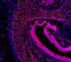
- Main image
- Experimental details
- FoxC2 in Mouse Embryo. FoxC2 was detected in immersion fixed frozen sections of mouse embryo (E15.5) using Sheep Anti-Mouse FoxC2 Antigen Affinity-purified Polyclonal Antibody (Catalog # AF6989) at 10 µg/mL overnight at 4 °C. Tissue was stained using the NorthernLights™ 557-conjugated Anti-Sheep IgG Secondary Antibody (red; Catalog # NL010) and counterstained with DAPI (blue). Specific staining was localized to periocular mesenchyme. View our protocol for Fluorescent IHC Staining of Frozen Tissue Sections.