Explore
 Validate
 Learn
Learn Immunohistochemistry
Antibody data
- Antibody Data
- Antigen structure
- References [4]
- Comments [0]
- Validations
- Immunohistochemistry [1]
Submit
Validation data
Reference
Comment
Report error
- Product number
- HPA017737 - Provider product page

- Provider
- Atlas Antibodies
- Proper citation
- Atlas Antibodies Cat#HPA017737, RRID:AB_1848107
- Product name
- Anti-PI3
- Antibody type
- Polyclonal
- Description
- Polyclonal Antibody against Human PI3, Gene description: peptidase inhibitor 3, skin-derived, Alternative Gene Names: cementoin, ELAFIN, ESI, SKALP, WAP3, WFDC14, Validated applications: IHC, Uniprot ID: P19957, Storage: Store at +4°C for short term storage. Long time storage is recommended at -20°C.
- Reactivity
- Human
- Host
- Rabbit
- Conjugate
- Unconjugated
- Isotype
- IgG
- Vial size
- 100 µl
- Concentration
- 0.1 mg/ml
- Storage
- Store at +4°C for short term storage. Long time storage is recommended at -20°C.
- Handling
- The antibody solution should be gently mixed before use.
Submitted references High circulating elafin levels are associated with Crohn’s disease-associated intestinal strictures
A High-throughput Bead-based Affinity Assay Enables Analysis of Genital Protein Signatures in Women At Risk of HIV Infection
Urinary Elafin and Kidney Injury in Hematopoietic Cell Transplant Recipients
Profiling post-centrifugation delay of serum and plasma with antibody bead arrays
Szecsi P, Wang J, Ortiz C, Fontenot L, Xie Y, Ho W, Mattai S, Shih D, Koon H
PLOS ONE 2020;15(4):e0231796
PLOS ONE 2020;15(4):e0231796
A High-throughput Bead-based Affinity Assay Enables Analysis of Genital Protein Signatures in Women At Risk of HIV Infection
Månberg A, Bradley F, Qundos U, Guthrie B, Birse K, Noël-Romas L, Lindskog C, Bosire R, Kiarie J, Farquhar C, Burgener A, Nilsson P, Broliden K
Molecular & Cellular Proteomics 2019;18(3):461-476
Molecular & Cellular Proteomics 2019;18(3):461-476
Urinary Elafin and Kidney Injury in Hematopoietic Cell Transplant Recipients
Hingorani S, Finn L, Pao E, Lawler R, Schoch G, McDonald G, Najafian B, Sandmaier B, Gooley T
Clinical Journal of the American Society of Nephrology 2015;10(1):12-20
Clinical Journal of the American Society of Nephrology 2015;10(1):12-20
Profiling post-centrifugation delay of serum and plasma with antibody bead arrays
Qundos U, Hong M, Tybring G, Divers M, Odeberg J, Uhlen M, Nilsson P, Schwenk J
Journal of Proteomics 2013;95
Journal of Proteomics 2013;95
No comments: Submit comment
Supportive validation
- Submitted by
- Atlas Antibodies (provider)
- Enhanced method
- Orthogonal validation
- Main image
- Experimental details
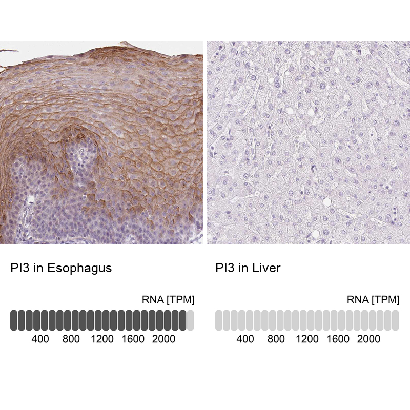
- Immunohistochemistry analysis in human esophagus and liver tissues using HPA017737 antibody. Corresponding PI3 RNA-seq data are presented for the same tissues.
- Sample type
- Human
- Protocol
- Protocol