 Explore
 Validate
 Learn
LearnHPA019947
antibody from Atlas Antibodies
Targeting: RUVBL1
ECP54, INO80H, NMP238, Pontin52, RVB1, TIH1, TIP49, TIP49a
 Western blot
 Immunocytochemistry
Antibody data
- Antibody Data
- Antigen structure
- References [4]
- Comments [0]
- Validations
- Western blot [1]
- Immunocytochemistry [1]
- Immunohistochemistry [1]
Submit
Validation data
Reference
Comment
Report error
- Product number
- HPA019947 - Provider product page

- Provider
- Atlas Antibodies
- Proper citation
- Atlas Antibodies Cat#HPA019947, RRID:AB_1856516
- Product name
- Anti-RUVBL1
- Antibody type
- Polyclonal
- Description
- Polyclonal Antibody against Human RUVBL1, Gene description: RuvB-like AAA ATPase 1, Alternative Gene Names: ECP54, INO80H, NMP238, Pontin52, RVB1, TIH1, TIP49, TIP49a, Validated applications: WB, IHC, ICC, Uniprot ID: Q9Y265, Storage: Store at +4°C for short term storage. Long time storage is recommended at -20°C.
- Reactivity
- Human
- Host
- Rabbit
- Conjugate
- Unconjugated
- Isotype
- IgG
- Vial size
- 100 µl
- Concentration
- 0.2 mg/ml
- Storage
- Store at +4°C for short term storage. Long time storage is recommended at -20°C.
- Handling
- The antibody solution should be gently mixed before use.
Submitted references Epigenetic Control of Translation Checkpoint and Tumor Progression via RUVBL1‐EEF1A1 Axis
Plasma cells in human pancreatic ductal adenocarcinoma secrete antibodies against self-antigens.
The relationship between RUVBL1 (Pontin, TIP49, NMP238) and BCL6 in benign and malignant human lymphoid tissues
Cancer Cell Response to Anthracyclines Effects: Mysteries of the Hidden Proteins Associated with These Drugs
Li M, Yang L, Chan A, Pokharel S, Liu Q, Mattson N, Xu X, Chang W, Miyashita K, Singh P, Zhang L, Li M, Wu J, Wang J, Chen B, Chan L, Lee J, Zhang X, Rosen S, Müschen M, Qi J, Chen J, Hiom K, Bishop A, Chen C
Advanced Science 2023;10(17)
Advanced Science 2023;10(17)
Plasma cells in human pancreatic ductal adenocarcinoma secrete antibodies against self-antigens.
Yao M, Preall J, Yeh JT, Pappin D, Cifani P, Zhao Y, Shen S, Moresco P, He B, Patel H, Habowski AN, King DA, Raphael K, Rishi A, Sejpal D, Weiss MJ, Tuveson D, Fearon DT
JCI insight 2023 Nov 8;8(21)
JCI insight 2023 Nov 8;8(21)
The relationship between RUVBL1 (Pontin, TIP49, NMP238) and BCL6 in benign and malignant human lymphoid tissues
Baron B, Baron R, Baron J
Biochemistry and Biophysics Reports 2016;6
Biochemistry and Biophysics Reports 2016;6
Cancer Cell Response to Anthracyclines Effects: Mysteries of the Hidden Proteins Associated with These Drugs
Tyleckova J, Hrabakova R, Mairychova K, Halada P, Radova L, Dzubak P, Hajduch M, Gadher S, Kovarova H
International Journal of Molecular Sciences 2012;13(12):15536-15564
International Journal of Molecular Sciences 2012;13(12):15536-15564
No comments: Submit comment
Enhanced validation
- Submitted by
- Atlas Antibodies (provider)
- Enhanced method
- Genetic validation
- Main image

- Experimental details
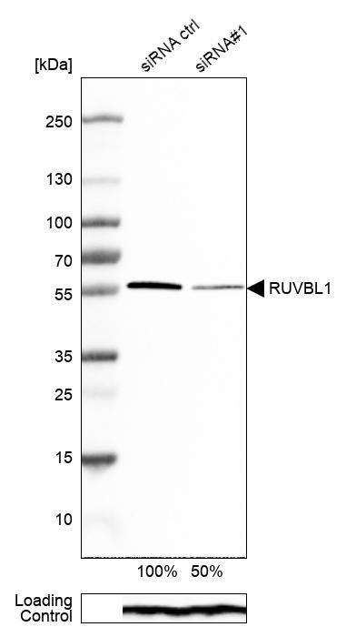
- Western blot analysis in MCF-7 cells transfected with control siRNA, target specific siRNA probe #1, using Anti-RUVBL1 antibody. Remaining relative intensity is presented. Loading control: Anti-PPIB.
- Sample type
- Human
- Protocol
- Protocol
Supportive validation
- Submitted by
- Atlas Antibodies (provider)
- Main image
- Experimental details
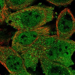
- Immunofluorescent staining of human cell line A-431 shows localization to nucleoplasm & cytosol.
- Sample type
- Human

Supportive validation
- Submitted by
- Atlas Antibodies (provider)
- Enhanced method
- Orthogonal validation
- Main image
- Experimental details
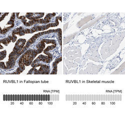
- Immunohistochemistry analysis in human fallopian tube and skeletal muscle tissues using Anti-RUVBL1 antibody. Corresponding RUVBL1 RNA-seq data are presented for the same tissues.
- Sample type
- Human
- Protocol
- Protocol
