Explore
 Validate
 Learn
LearnPA5-29278
antibody from Invitrogen Antibodies
Targeting: RUVBL1
ECP54, INO80H, NMP238, Pontin52, RVB1, TIH1, TIP49, TIP49a
 Western blot
 Immunocytochemistry
Antibody data
- Antibody Data
- Antigen structure
- References [2]
- Comments [0]
- Validations
- Immunocytochemistry [3]
- Immunoprecipitation [1]
- Immunohistochemistry [5]
- Chromatin Immunoprecipitation [2]
- Other assay [1]
Submit
Validation data
Reference
Comment
Report error
- Product number
- PA5-29278 - Provider product page

- Provider
- Invitrogen Antibodies
- Product name
- RUVBL1 Polyclonal Antibody
- Antibody type
- Polyclonal
- Antigen
- Recombinant full-length protein
- Description
- Recommended positive controls: A549, H1299, HCT116. Predicted reactivity: Mouse (99%), Rat (99%), Zebrafish (97%), Xenopus laevis (97%), Chicken (99%), Rhesus Monkey (100%), Chimpanzee (100%), Bovine (99%). Store product as a concentrated solution. Centrifuge briefly prior to opening the vial.
- Reactivity
- Human, Mouse, Rat
- Host
- Rabbit
- Isotype
- IgG
- Vial size
- 100 μL
- Concentration
- 1.98 mg/mL
- Storage
- Store at 4°C short term. For long term storage, store at -20°C, avoiding freeze/thaw cycles.
Submitted references RuvBL1 Maintains Resistance to TRAIL-Induced Apoptosis by Suppressing c-Jun/AP-1 Activity in Non-Small Cell Lung Cancer.
PRMT5-Dependent Methylation of the TIP60 Coactivator RUVBL1 Is a Key Regulator of Homologous Recombination.
Li H, Zhou T, Zhang Y, Jiang H, Zhang J, Hua Z
Frontiers in oncology 2021;11:679243
Frontiers in oncology 2021;11:679243
PRMT5-Dependent Methylation of the TIP60 Coactivator RUVBL1 Is a Key Regulator of Homologous Recombination.
Clarke TL, Sanchez-Bailon MP, Chiang K, Reynolds JJ, Herrero-Ruiz J, Bandeiras TM, Matias PM, Maslen SL, Skehel JM, Stewart GS, Davies CC
Molecular cell 2017 Mar 2;65(5):900-916.e7
Molecular cell 2017 Mar 2;65(5):900-916.e7
No comments: Submit comment
Supportive validation
- Submitted by
- Invitrogen Antibodies (provider)
- Main image
- Experimental details
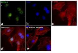
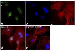
- Immunofluorescence analysis of RUVBL1 was performed using 70% confluent log phase A549 cells. The cells were fixed with 4% paraformaldehyde for 10 minutes, permeabilized with 0.1% Triton™ X-100 for 10 minutes, and blocked with 1% BSA for 1 hour at room temperature. The cells were labeled with RUVBL1 Rabbit Polyclonal Antibody (Product # PA5-29278) at 5 µg/mL in 0.1% BSA and incubated overnight at 4 degree and then labeled with Goat anti-Rabbit IgG (H+L) Superclonal™ Secondary Antibody, Alexa Fluor® 488 conjugate (Product # A27034) at a dilution of 1:2000 for 45 minutes at room temperature (Panel a: green). Nuclei (Panel b: blue) were stained with SlowFade® Gold Antifade Mountant with DAPI (Product # S36938). F-actin (Panel c: red) was stained with Rhodamine Phalloidin (Product # R415, 1:300). Panel d represents the merged image showing nuclear localization. Panel e represents control cells with no primary antibody to assess background. The images were captured at 60X magnification.
- Submitted by
- Invitrogen Antibodies (provider)
- Main image
- Experimental details
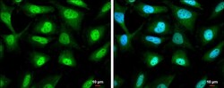
- Immunocytochemistry-Immunofluorescence analysis of RUVBL1 was performed in HeLa cells fixed in 4% paraformaldehyde at RT for 15 min. Green: RUVBL1 Polyclonal Antibody (Product # PA5-29278) diluted at 1:500. Blue: Hoechst 33342 staining. Scale bar = 10 µm.

- Submitted by
- Invitrogen Antibodies (provider)
- Main image
- Experimental details
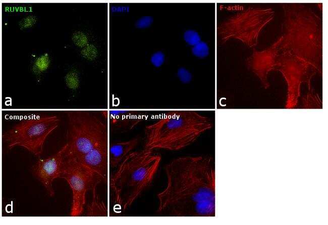
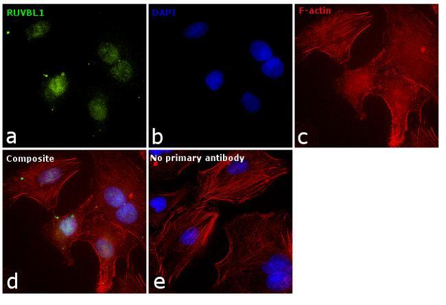
- Immunofluorescence analysis of RUVBL1 was performed using 70% confluent log phase A549 cells. The cells were fixed with 4% paraformaldehyde for 10 minutes, permeabilized with 0.1% Triton™ X-100 for 10 minutes, and blocked with 1% BSA for 1 hour at room temperature. The cells were labeled with RUVBL1 Rabbit Polyclonal Antibody (Product # PA5-29278) at 5 µg/mL in 0.1% BSA and incubated overnight at 4 degree and then labeled with Goat anti-Rabbit IgG (Heavy Chain) Superclonal™ Secondary Antibody, Alexa Fluor® 488 conjugate (Product # A27034) at a dilution of 1:2000 for 45 minutes at room temperature (Panel a: green). Nuclei (Panel b: blue) were stained with SlowFade® Gold Antifade Mountant with DAPI (Product # S36938). F-actin (Panel c: red) was stained with Rhodamine Phalloidin (Product # R415, 1:300). Panel d represents the merged image showing nuclear localization. Panel e represents control cells with no primary antibody to assess background. The images were captured at 60X magnification.
Supportive validation
- Submitted by
- Invitrogen Antibodies (provider)
- Main image
- Experimental details
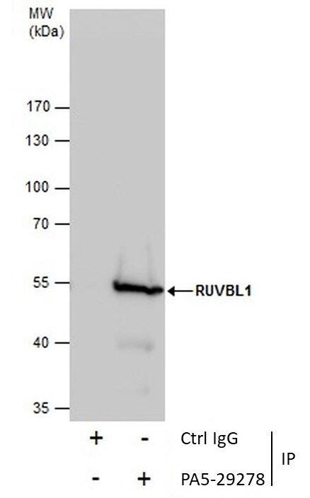
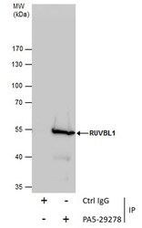
- Immunoprecipitation of RUVBL was performed in Jurkat whole cell extracts using 5 µg of RUVBL1 Polyclonal Antibody (Product # PA5-29278). Samples were transferred to a membrane and probed with RUVBL1 Polyclonal Antibody as a primary antibody and an HRP-conjugated anti-Rabbit IgG was used as a secondary antibody.

Supportive validation
- Submitted by
- Invitrogen Antibodies (provider)
- Main image

- Experimental details
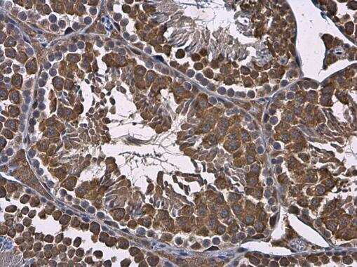
- Immunohistochemistry (Paraffin) analysis of RUVBL1 was performed in paraffin-embedded mouse testis tissue using RUVBL1 Polyclonal Antibody (Product # PA5-29278) at a dilution of 1:500.
- Submitted by
- Invitrogen Antibodies (provider)
- Main image
- Experimental details
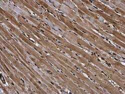
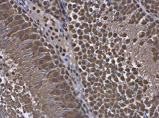
- Immunohistochemistry (Paraffin) analysis of RUVBL1 was performed in paraffin-embedded rat heart tissue using RUVBL1 Polyclonal Antibody (Product # PA5-29278) at a dilution of 1:500.

- Submitted by
- Invitrogen Antibodies (provider)
- Main image
- Experimental details
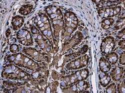
- Immunohistochemistry (Paraffin) analysis of RUVBL1 was performed in paraffin-embedded mouse colon tissue using RUVBL1 Polyclonal Antibody (Product # PA5-29278) at a dilution of 1:500.
- Submitted by
- Invitrogen Antibodies (provider)
- Main image
- Experimental details
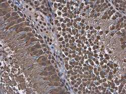
- Immunohistochemistry (Paraffin) analysis of RUVBL1 was performed in paraffin-embedded rat testis tissue using RUVBL1 Polyclonal Antibody (Product # PA5-29278) at a dilution of 1:500.
- Submitted by
- Invitrogen Antibodies (provider)
- Main image

- Experimental details
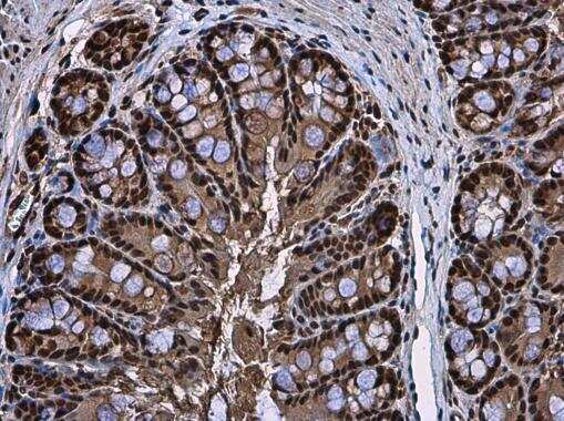
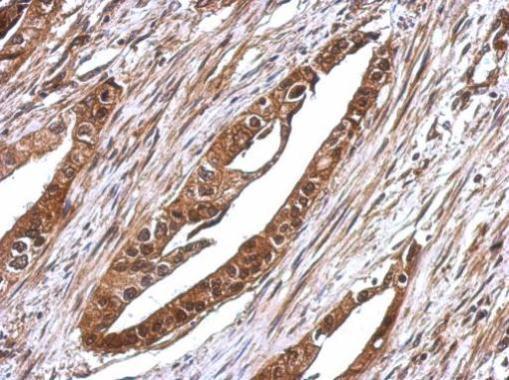
- Immunohistochemical analysis of paraffin-embedded human colon carcinoma, using RUVBL1 (Product # PA5-29278) antibody at 1:500 dilution. Antigen Retrieval: EDTA based buffer, pH 8.0, 15 min.
Supportive validation
- Submitted by
- Invitrogen Antibodies (provider)
- Main image

- Experimental details
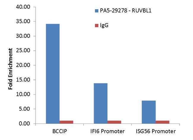
- Enrichment of endogenous RUVBL1 protein at specific gene loci using Anti-RUVBL1 Antibody: Chromatin Immunoprecipitation (ChIP) was performed using Anti-RUVBL1 Rabbit Polyclonal Antibody (Product # PA5-29278, 4 µg) on sheared chromatin from 2 million SW480 cells using the MAGnify ChIP system kit (Product # 49-2024). Normal Rabbit IgG was used as a negative IP control. The purified DNA was analyzed by qPCR with PCR primer pairs over the BCCIP gene and promoters of IFI6 and ISG56. Data is presented as fold enrichment of the antibody signal versus the negative control IgG using the comparative CT method.
- Submitted by
- Invitrogen Antibodies (provider)
- Main image

- Experimental details
- Enrichment of endogenous RUVBL1 protein at specific gene loci using Anti-RUVBL1 Antibody: Chromatin Immunoprecipitation (ChIP) was performed using Anti-RUVBL1 Rabbit Polyclonal Antibody (Product # PA5-29278, 4 µg) on sheared chromatin from 2 million SW480 cells using the MAGnify ChIP system kit (Product # 49-2024). Normal Rabbit IgG was used as a negative IP control. The purified DNA was analyzed by qPCR with PCR primer pairs over the BCCIP gene and promoters of IFI6 and ISG56. Data is presented as fold enrichment of the antibody signal versus the negative control IgG using the comparative CT method.

Supportive validation
- Submitted by
- Invitrogen Antibodies (provider)
- Main image

- Experimental details
- Immunoprecipitation of RUVBL was performed in Jurkat whole cell extracts using 5 µg of RUVBL1 Polyclonal Antibody (Product # PA5-29278). Samples were transferred to a membrane and probed with RUVBL1 Polyclonal Antibody as a primary antibody and an HRP-conjugated anti-Rabbit IgG was used as a secondary antibody.