Explore
 Validate
 Learn
Learn Immunocytochemistry
Antibody data
- Antibody Data
- Antigen structure
- References [1]
- Comments [0]
- Validations
- Immunocytochemistry [1]
- Other assay [1]
Submit
Validation data
Reference
Comment
Report error
- Product number
- PA5-47565 - Provider product page

- Provider
- Invitrogen Antibodies
- Product name
- MYF5 Polyclonal Antibody
- Antibody type
- Polyclonal
- Antigen
- Recombinant full-length protein
- Description
- This antibody detects recombinant human MYF-5 in direct ELISAs and Western blots. Reconstitute at 0.2 mg/mL in sterile PBS.
- Reactivity
- Human, Mouse
- Host
- Goat
- Isotype
- IgG
- Vial size
- 100 μg
- Concentration
- 0.2 mg/mL
- Storage
- -20°C, Avoid Freeze/Thaw Cycles
Submitted references Treatment with galectin-1 improves myogenic potential and membrane repair in dysferlin-deficient models.
Vallecillo-Zúniga ML, Rathgeber MF, Poulson PD, Hayes S, Luddington JS, Gill HN, Teynor M, Kartchner BC, Valdoz J, Stowell C, Markham AR, Arthur C, Stowell S, Van Ry PM
PloS one 2020;15(9):e0238441
PloS one 2020;15(9):e0238441
No comments: Submit comment
Supportive validation
- Submitted by
- Invitrogen Antibodies (provider)
- Main image
- Experimental details
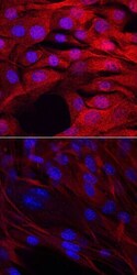
- Immunocytochemistry analysis of MYF5 in immersion fixed C2C12 mouse myoblast cell line undifferentiated (upper panel) or differentiated with horse serum for 7 days (lower panel). Samples were incubated in MYF5 polyclonal antibody (Product # PA5-47565) using a dilution of 10 µg/mL for 3 hours at room temperature followed by NorthernLights™ 557-conjugated Anti-Goat IgG Secondary Antibody (red) and counterstained with DAPI (blue).

Supportive validation
- Submitted by
- Invitrogen Antibodies (provider)
- Main image
- Experimental details
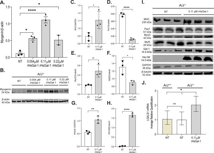
- Fig 1 rHsGal-1 increases myogenic regulatory factors in A/J -/- myotubes. A. Quantification of myogenin after 72h treatment with varying concentrations of rHsGal-1. B. Western blot images of myogenin at different rHsGal-1 treatments. C-F. Quantification of myogenic markers MHC(C), Pax7(D), MyoD(E), and Myf5(F) in A/J -/- myotubes after 72h treatment with 0.11muM rHsGal-1. G-H. Quantification of Gal-1(G) and His.H8(H)in A/J -/- myotubes after 72h treatment with 0.11muM rHsGal-1. I. Western blot images of myogenic markers (Pax7, Myf5, MyoD, and MHC) and of mouse Gal-1 and His Tagged rHsGal-1. J. RT-qPCR quantification of LGALS1 transcript between A/J WT, A/J -/- NT, and A/J -/- 0.11muM rHsGal-1 treated myotubes. p values are measured by Tukey's multiple comparison test and indicated by *p< 0.05, **p< 0.01, ****p< 0.0001 (n = 3 for each group). Error bars represent SEM.