 Explore
 Validate
 Learn
Learn Western blot
Antibody data
- Antibody Data
- Antigen structure
- References [0]
- Comments [0]
- Validations
- Western blot [1]
- Immunohistochemistry [1]
Submit
Validation data
Reference
Comment
Report error
- Product number
- MAB7627-100 - Provider product page

- Provider
- R&D Systems
- Product name
- Human Prostaglandin E Synthase 2/PTGES2 Antibody
- Antibody type
- Monoclonal
- Description
- Protein A or G purified from cell culture supernatant. Detects Human Prostaglandin E Synthase 2/PTGES2 in direct ELISAs.
- Reactivity
- Human
- Host
- Mouse
- Conjugate
- Unconjugated
- Antigen sequence
Q9H7Z7- Isotype
- IgG
- Antibody clone number
- 998012
- Vial size
- 100 ug
- Storage
- Use a manual defrost freezer and avoid repeated freeze-thaw cycles. 12 months from date of receipt, -20 to -70 °C as supplied. 1 month, 2 to 8 °C under sterile conditions after reconstitution. 6 months, -20 to -70 °C under sterile conditions after reconstitution.
No comments: Submit comment
Supportive validation
- Submitted by
- R&D Systems (provider)
- Main image

- Experimental details
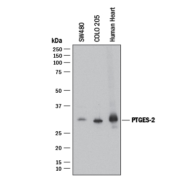
- Detection of Human Prostaglandin E Synthase 2/PTGES2 by Western Blot. Western blot shows lysates of SW480 human colorectal adenocarcinoma cell line, COLO 205 human colorectal adenocarcinoma cell line, and human heart tissue. PVDF membrane was probed with 2 µg/mL of Mouse Anti-Human Prostaglandin E Synthase 2/PTGES2 Monoclonal Antibody (Catalog # MAB7627) followed by HRP-conjugated Anti-Mouse IgG Secondary Antibody (Catalog # HAF018). A specific band was detected for Prostaglandin E Synthase 2/PTGES2 at approximately 30 kDa (as indicated). This experiment was conducted under reducing conditions and using Immunoblot Buffer Group 1.
Supportive validation
- Submitted by
- R&D Systems (provider)
- Main image
- Experimental details
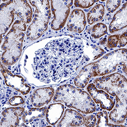
- Prostaglandin E Synthase 2/PTGES2 in Human Kidney. Prostaglandin E Synthase 2/PTGES2 was detected in immersion fixed paraffin-embedded sections of human kidney using Mouse Anti-Human Prostaglandin E Synthase 2/PTGES2 Monoclonal Antibody (Catalog # MAB7627) at 5 µg/mL for 1 hour at room temperature followed by incubation with the Anti-Mouse IgG VisUCyte™ HRP Polymer Antibody (Catalog # VC001). Before incubation with the primary antibody, tissue was subjected to heat-induced epitope retrieval using Antigen Retrieval Reagent-Basic (Catalog # CTS013). Tissue was stained using DAB (brown) and counterstained with hematoxylin (blue). Specific staining was localized to cytoplasm in epithelial cells in convoluted tubules. View our protocol for IHC Staining with VisUCyte HRP Polymer Detection Reagents.
