Explore
 Validate
 Learn
Learn Western blot
 Immunocytochemistry
Antibody data
- Antibody Data
- Antigen structure
- References [2]
- Comments [0]
- Validations
- Immunocytochemistry [2]
- Immunohistochemistry [1]
- Flow cytometry [1]
Submit
Validation data
Reference
Comment
Report error
- Product number
- 700180 - Provider product page

- Provider
- Invitrogen Antibodies
- Product name
- PSME3 Recombinant Rabbit Monoclonal Antibody (20H9L19)
- Antibody type
- Monoclonal
- Antigen
- Synthetic peptide
- Description
- This antibody is predicted to react with canine, chicken, equine, orangutan, Xenopus and zebrafish based on sequence homology. Intact IgG appears on a non-reducing gel as ~150 kDa band and upon reduction generating a ~25 kDa light chain band and a ~50 kDa heavy chain. Recombinant rabbit monoclonal antibodies are produced using in vitro expression systems. The expression systems are developed by cloning in the specific antibody DNA sequences from immunoreactive rabbits. Then, individual clones are screened to select the best candidates for production. The advantages of using recombinant rabbit monoclonal antibodies include: better specificity and sensitivity, lot-to-lot consistency, animal origin-free formulations, and broader immunoreactivity to diverse targets due to larger rabbit immune repertoire.
- Reactivity
- Human, Mouse, Rat
- Host
- Rabbit
- Isotype
- IgG
- Antibody clone number
- 20H9L19
- Vial size
- 100 μg
- Concentration
- 0.5 mg/mL
- Storage
- Store at 4°C short term. For long term storage, store at -20°C, avoiding freeze/thaw cycles.
Submitted references Site-specific O-GlcNAcylation of Psme3 maintains mouse stem cell pluripotency by impairing P-body homeostasis.
Mutant p53 (p53-R248Q) functions as an oncogene in promoting endometrial cancer by up-regulating REGγ.
Pecori F, Kondo N, Ogura C, Miura T, Kume M, Minamijima Y, Yamamoto K, Nishihara S
Cell reports 2021 Jul 13;36(2):109361
Cell reports 2021 Jul 13;36(2):109361
Mutant p53 (p53-R248Q) functions as an oncogene in promoting endometrial cancer by up-regulating REGγ.
Wang H, Bao W, Jiang F, Che Q, Chen Z, Wang F, Tong H, Dai C, He X, Liao Y, Liu B, Sun J, Wan X
Cancer letters 2015 May 1;360(2):269-79
Cancer letters 2015 May 1;360(2):269-79
No comments: Submit comment
Supportive validation
- Submitted by
- Invitrogen Antibodies (provider)
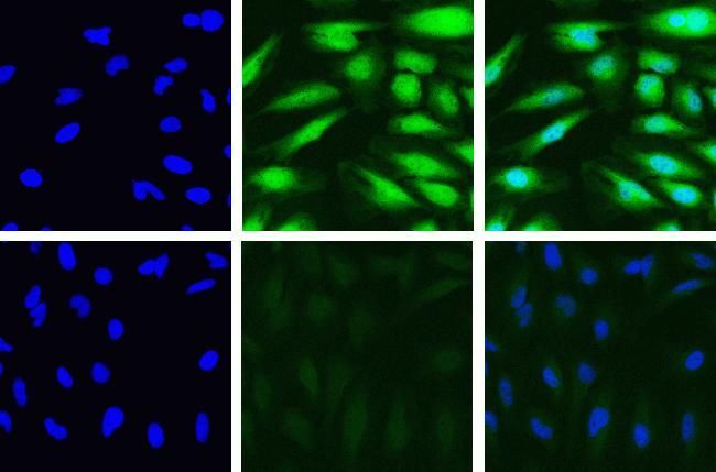
- Main image
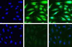
- Experimental details
- Immunofluorescent analysis of PSME3 in HeLa cells using a PSME3 recombinant rabbit monoclonal antibody (Product # 700180) at a dilution of 5 µg/mL in the absence of peptide (top) and presence of immunogenic peptide (bottom), followed by detection using an Alexa Fluor 488-conjugated goat anti-rabbit secondary antibody at a dilution of 1:1000. Actin was stained with Alexa Fluor 568 Phalloidin (Product # A12380). Hoechst only (blue, left), AF488 signal only (green, middle) and composite image with Phalloidin (right).

- Submitted by
- Invitrogen Antibodies (provider)
- Main image
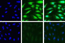
- Experimental details
- Immunofluorescent analysis of PSME3 in HeLa cells using a PSME3 recombinant rabbit monoclonal antibody (Product # 700180) at a dilution of 5 µg/mL in the absence of peptide (top) and presence of immunogenic peptide (bottom), followed by detection using an Alexa Fluor 488-conjugated goat anti-rabbit secondary antibody at a dilution of 1:1000. Actin was stained with Alexa Fluor 568 Phalloidin (Product # A12380). Hoechst only (blue, left), AF488 signal only (green, middle) and composite image with Phalloidin (right).
Supportive validation
- Submitted by
- Invitrogen Antibodies (provider)
- Main image
- Experimental details
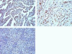
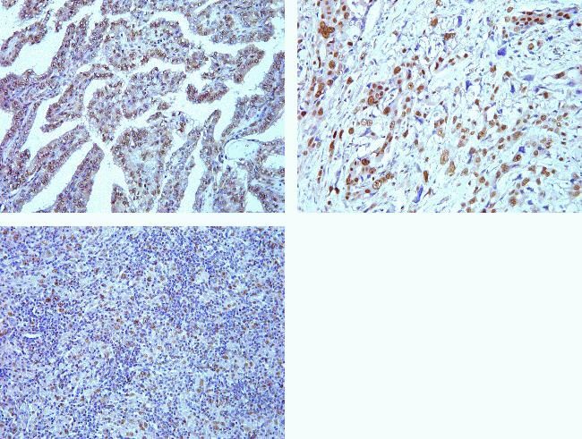
- Immunohistochemistry analysis of PSME3 in formalin-fixed, paraffin-embedded human thyroid (top left) and lung carcimona (top right) and Hodgkin lymphoma (bottom left) using a PSME3 monoclonal antibody (Product # 700180) at a dilution of 5 µg/mL. Staining was visualized using DAB and images were taken at a magnification of 20x. Results show strong nuclear staining in tumor cells.
Supportive validation
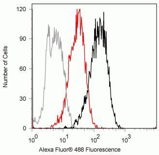
- Submitted by
- Invitrogen Antibodies (provider)
- Main image

- Experimental details
- Flow cytometry analysis of PSME3 in Jurkat cells using a PSME3 recombinant rabbit monoclonal antibody (Product # 700180) at a dilution of 2ug. Cells were fixed and permeabilized using FIX & PERM (Product # GAS004) reagent, and detection was performed using an Alexa Fluor 488 goat anti-rabbit IgG (black) compared to a control without primary antibody (gray). Pre-incubation with the immunogenic peptide decreased the signal (red).