Explore
 Validate
 Learn
Learn Western blot
 Immunoprecipitation
 Chromatin Immunoprecipitation
Antibody data
- Antibody Data
- Antigen structure
- References [0]
- Comments [0]
- Validations
- Chromatin Immunoprecipitation [2]
- Other assay [1]
Submit
Validation data
Reference
Comment
Report error
- Product number
- A303-381A - Provider product page

- Provider
- Invitrogen Antibodies
- Product name
- IRF2 Polyclonal Antibody
- Antibody type
- Polyclonal
- Antigen
- Other
- Reactivity
- Human
- Host
- Rabbit
- Isotype
- IgG
- Vial size
- 100 µL
- Concentration
- 1 mg/mL
- Storage
- 4° C
No comments: Submit comment
Supportive validation
- Submitted by
- Invitrogen Antibodies (provider)
- Main image
- Experimental details
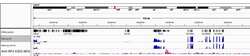
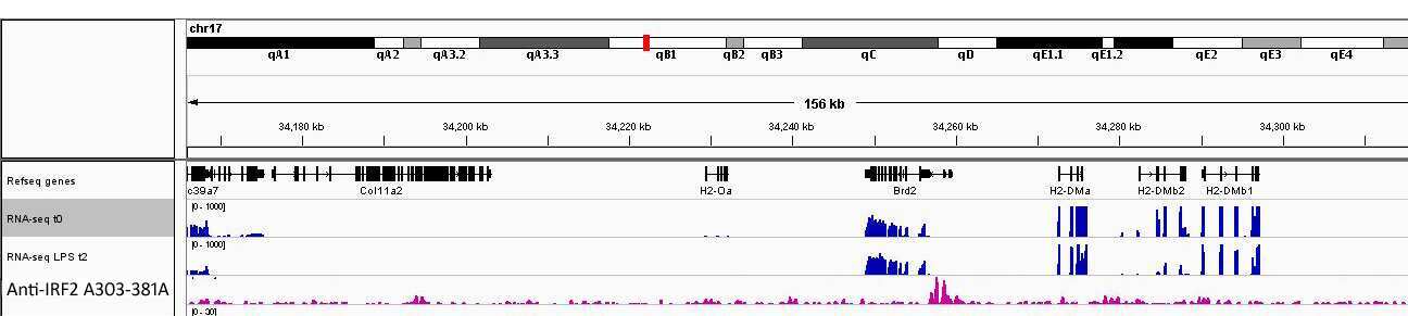
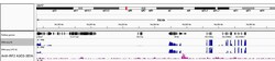
- Localization of IRF2 binding sites by ChIP-Sequencing. Chromatin from mouse bone marrow dendritic cells (BMDCs) was immunoprecipitated with anti-IRF2 A303-381A (lot A303-381A-1) and analyzed by DNA sequencing according to methods available at http://www.weizmann.ac.il/immunology/AmitLab/data-and-method/iChIP/method. The figure illustrates the peak distribution of IRF2 binding within a 156 kb region of chromosome 17 (pink) as detected by anti-IRF2 A303-381A.
- Submitted by
- Invitrogen Antibodies (provider)
- Main image
- Experimental details
- Localization of IRF2 binding sites by ChIP-Sequencing. Chromatin from mouse bone marrow dendritic cells (BMDCs) was immunoprecipitated with anti-IRF2 A303-381A (lot A303-381A-1) and analyzed by DNA sequencing according to methods available at http://www.weizmann.ac.il/immunology/AmitLab/data-and-method/iChIP/method. The figure illustrates the peak distribution of IRF2 binding within a 156 kb region of chromosome 17 (pink) as detected by anti-IRF2 A303-381A.

Supportive validation
- Submitted by
- Invitrogen Antibodies (provider)
- Main image

- Experimental details
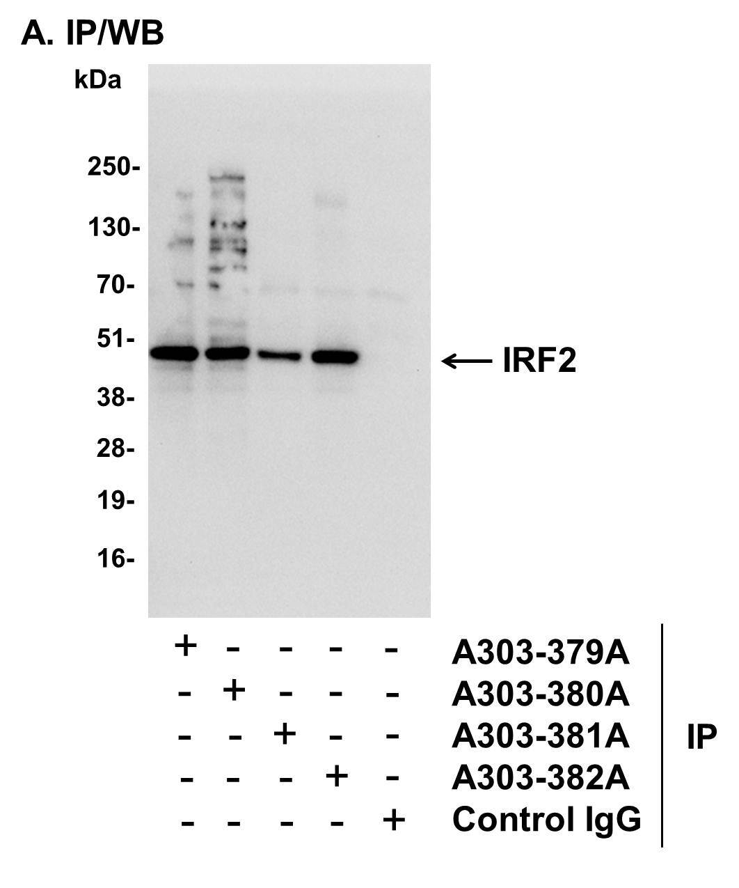
- Detection of human IRF2 by western blot of immunoprecipitates. Samples: Whole cell lysate (1 mg for IP, 20% of IP loaded) from human erythroleukemia (HEL) cells. Antibodies: Affinity purified rabbit anti-IRF2 antibody A303-381A used for IP at 6 µg/mg lysate. IRF2 was also immunoprecipitated by rabbit anti-IRF2 antibodies A303-379A , A303-380A, and A303-382A, which recognize disparate epitopes. For blotting immunoprecipitated IRF2, A303-380A was used at 1 µg/ml. Detection: Chemiluminescence with an exposure time of 3 seconds.