Explore
 Validate
 Learn
Learn Western blot
 ELISA
ELISAAntibody data
- Antibody Data
- Antigen structure
- References [1]
- Comments [0]
- Validations
- Western blot [1]
- Immunohistochemistry [2]
Submit
Validation data
Reference
Comment
Report error
- Product number
- NBP1-77938 - Provider product page

- Provider
- Novus Biologicals
- Proper citation
- Novus Cat#NBP1-77938, RRID:AB_11037359
- Product name
- Rabbit Polyclonal MLF1 Interacting Protein Antibody
- Antibody type
- Polyclonal
- Description
- Immunogen affinity purified. This antibody is specific for human Mlf1 phospho T78 and surrounding amino acids.
- Reactivity
- Human, Bovine, Canine
- Host
- Rabbit
- Isotype
- IgG
- Vial size
- 0.1 mg
- Concentration
- 1 mg/ml
- Storage
- Store at -20C. Avoid freeze-thaw cycles.
Submitted references Self-regulated Plk1 recruitment to kinetochores by the Plk1-PBIP1 interaction is critical for proper chromosome segregation.
Kang YH, Park JE, Yu LR, Soung NK, Yun SM, Bang JK, Seong YS, Yu H, Garfield S, Veenstra TD, Lee KS
Molecular cell 2006 Nov 3;24(3):409-22
Molecular cell 2006 Nov 3;24(3):409-22
No comments: Submit comment
Supportive validation
- Submitted by
- Novus Biologicals (provider)
- Main image
- Experimental details
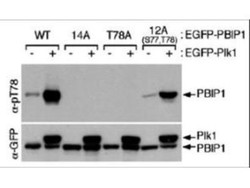
- Western Blot: MLF1 Interacting Protein [p Thr78] Antibody [NBP1-77938] - Shows detection of MLF1IP phosphorylated at Thr78. HeLa cells were co-infected with the indicated adenoviruses expressing GFP-tagged Plk1 or PBIP1. Blots were probed with the anti-MLF1IP pT78 antibody, stripped, and then reprobed with anti-GFP antibody (KA/G & Park, et al., 2006).

Supportive validation
- Submitted by
- Novus Biologicals (provider)
- Main image
- Experimental details
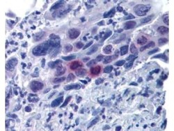
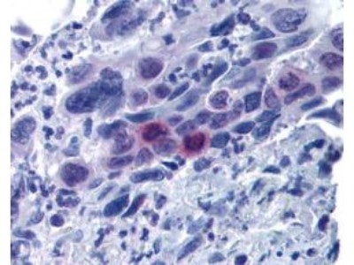
- Immunohistochemistry: MLF1 Interacting Protein [p Thr78] Antibody [NBP1-77938] - Used at 20 g/ml to detect signal in a variety of tissues including multi-human, multi-brain and multi-cancer slides. This image shows moderately positive staining of mitotic cells in colon adenocarcinoma at 60X. Tissue was formalin-fixed and paraffin embedded. The image shows localization of the antibody as the precipitated red signal, with a hematoxylin purple nuclear counterstain. Personal Communication, Tina Roush, LifeSpanBiosciences, Seattle, WA.
- Submitted by
- Novus Biologicals (provider)
- Main image

- Experimental details
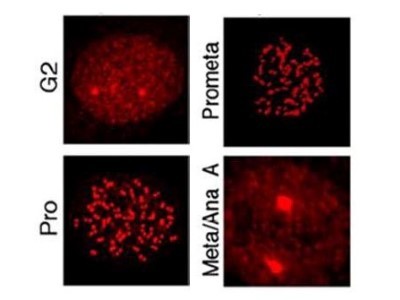
- Immunohistochemistry: MLF1 Interacting Protein [p Thr78] Antibody [NBP1-77938] - Analysis of MLF1IP pT78 at the kinetochores of HeLa cells in different phases of the cell cycle. Fluorescent signals were detectable at the kinetochores as early as G2, became most abundant in prophase cells with a discernible nuclear envelope, and gradually diminished as cells proceeded through mitosis (Kang & Park, et al., 2006).