Explore
 Validate
 Learn
LearnAP09322PU-N
antibody from Acris Antibodies GmbH
Targeting: CENPU
CENP-50, CENP-U, KLIP1, MLF1IP, PBIP1
 Western blot
 ELISA
ELISAAntibody data
- Antibody Data
- Antigen structure
- References [0]
- Comments [0]
- Validations
- Western blot [1]
- Immunocytochemistry [1]
- Immunohistochemistry [1]
Submit
Validation data
Reference
Comment
Report error
- Product number
- AP09322PU-N - Provider product page

- Provider
- Acris Antibodies GmbH
- Proper citation
- Acris Antibodies GmbH Cat#AP09322PU-N, RRID:AB_2035771
- Product name
- anti MLF1IP pThr78
- Antibody type
- Polyclonal
- Antigen
- Synthetic peptide corresponding to amino acids surrounding Thr78 of human MLF1IP protein. The immunogen peptide is phosphorylated at Thr78
- Reactivity
- Human, Bovine, Canine
- Host
- Rabbit
- Isotype
- IgG
- Vial size
- 0.1 mg
- Concentration
- 1.26 mg/ml (by UV absorbance at 280 nm)
No comments: Submit comment
Supportive validation
- Submitted by
- Acris Antibodies GmbH (provider)
- Main image
- Experimental details
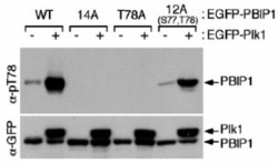
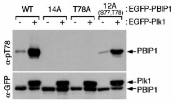
- Western blot using affinity purified anti-MLF1IP pT78 antibody shows detection of MLF1IP phosphorylated at Thr78. HeLa cells were co-infected with the indicated adenoviruses expressing GFP-tagged Plk1 or PBIP1. Blots were probed with the anti-MLF1IP pT78 antibody, stripped, and then reprobed with anti-GFP antibody (Kang & Park, et al., 2006).
Supportive validation
- Submitted by
- Acris Antibodies GmbH (provider)
- Main image
- Experimental details
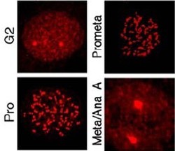
- Immunostaining using affinity purified anti-MLF1IP pT78 antibody shows detection of MLF1IP pT78 at the kinetochores of HeLa cells in different phases of the cell cycle. Fluorescent signals were detectable at the kinetochores as early as G2, became most abundant in prophase cells with a discernible nuclear envelope, and gradually diminished as cells proceeded through mitosis (Kang & Park, et al., 2006)

Supportive validation
- Submitted by
- Acris Antibodies GmbH (provider)
- Main image
- Experimental details
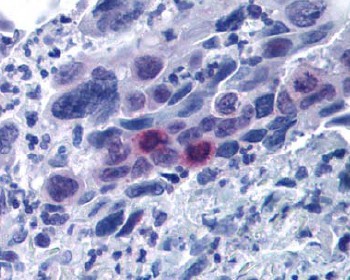
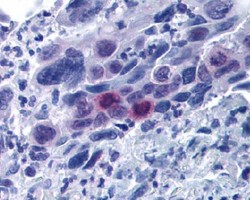
- Immunohistochemistry affinity purified anti-MLF1IP pT78 antibody was used at 20 µg/ml to detect signal in a variety of tissues including multi-human, multi-brain and multicancer slides. This image shows moderately positive staining of mitotic cells in colon adenocarcinoma at 60X. Tissue was formalin-fixed and paraffin embedded. The image shows localization of the antibody as the precipitated red signal, with a hematoxylin purple nuclear counterstain.