 Explore
 Validate
 Learn
Learn Western blot
 Immunocytochemistry
Antibody data
- Antibody Data
- Antigen structure
- References [1]
- Comments [0]
- Validations
- Immunocytochemistry [2]
- Chromatin Immunoprecipitation [2]
Submit
Validation data
Reference
Comment
Report error
- Product number
- 703747 - Provider product page

- Provider
- Invitrogen Antibodies
- Product name
- ZNF207 Recombinant Rabbit Monoclonal Antibody (7H6L2)
- Antibody type
- Monoclonal
- Antigen
- Other
- Description
- This antibody is predicted to react with Bovine.
- Reactivity
- Human, Mouse
- Host
- Rabbit
- Isotype
- IgG
- Antibody clone number
- 7H6L2
- Vial size
- 100 μg
- Concentration
- 0.5 mg/mL
- Storage
- Store at 4°C short term. For long term storage, store at -20°C, avoiding freeze/thaw cycles.
Submitted references System analysis based on the cancer-immunity cycle identifies ZNF207 as a novel immunotherapy target for hepatocellular carcinoma.
Wang X, Zhou T, Chen X, Wang Y, Ding Y, Tu H, Gao S, Wang H, Tang X, Yang Y
Journal for immunotherapy of cancer 2022 Mar;10(3)
Journal for immunotherapy of cancer 2022 Mar;10(3)
No comments: Submit comment
Supportive validation
- Submitted by
- Invitrogen Antibodies (provider)
- Main image
- Experimental details
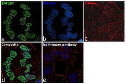
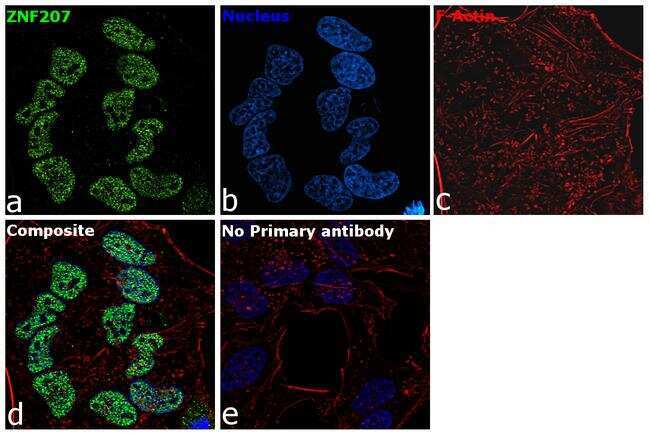
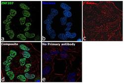
- For immunocytochemistry analysis, HeLa cells were fixed and permeabilized for detection of endogenous ZNF207 using Anti-ZNF207 Recombinant Rabbit Monoclonal Antibody (Product # 703747) at a 1:100 dilution and labeled with Goat anti-Rabbit IgG (H+L) Highly Cross-Adsorbed Secondary Antibody, Alexa Fluor Plus 488 conjugate (Product # A32731) at a 1:2000 dilution. Panel a) shows representative cells that were stained for detection and localization of ZNF207 protein (green), Panel b) is stained for nuclei (blue) using ProLong™ Diamond Antifade Mountant with DAPI (Product # P36962). Panel c) represents cytoskeletal F-actin staining using Rhodamine Phalloidin (Product # R415) at a 1:300 dilution. Panel d) is a composite image of Panels a, b and c clearly demonstrating nuclear localization of ZNF207. Panel e) represents control cells with no primary antibody to assess background. The images were captured at 60X magnification.
- Submitted by
- Invitrogen Antibodies (provider)
- Main image
- Experimental details
- For immunocytochemistry analysis, HeLa cells were fixed and permeabilized for detection of endogenous ZNF207 using Anti-ZNF207 Recombinant Rabbit Monoclonal Antibody (Product # 703747) at a 1:100 dilution and labeled with Goat anti-Rabbit IgG (H+L) Highly Cross-Adsorbed Secondary Antibody, Alexa Fluor Plus 488 conjugate (Product # A32731) at a 1:2000 dilution. Panel a) shows representative cells that were stained for detection and localization of ZNF207 protein (green), Panel b) is stained for nuclei (blue) using ProLong™ Diamond Antifade Mountant with DAPI (Product # P36962). Panel c) represents cytoskeletal F-actin staining using Rhodamine Phalloidin (Product # R415) at a 1:300 dilution. Panel d) is a composite image of Panels a, b and c clearly demonstrating nuclear localization of ZNF207. Panel e) represents control cells with no primary antibody to assess background. The images were captured at 60X magnification.

Supportive validation
- Submitted by
- Invitrogen Antibodies (provider)
- Main image

- Experimental details
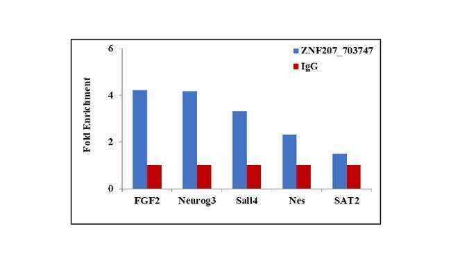
- Chromatin Immunoprecipitation (ChIP) assay of endogenous ZNF207 protein using Anti-ZNF207 Antibody: ChIP was performed using 5 µg of Anti-ZNF207 Recombinant Rabbit Monoclonal Antibody (Product # 703747) on sheared chromatin from 2 million HeLa cells using the MAGnify ChIP System kit (Product # 49-2024). Normal Rabbit IgG was used as a negative IP control. The purified DNA was analyzed by qPCR using primers binding to the transcriptional start site of FGF2, Neurog3, Sall4, Nes and SAT2 satellite repeats. Data is presented as fold enrichment of the antibody signal versus the negative control IgG using the comparative CT method.
- Submitted by
- Invitrogen Antibodies (provider)
- Main image

- Experimental details
- Chromatin Immunoprecipitation (ChIP) assay of endogenous ZNF207 protein using Anti-ZNF207 Antibody: ChIP was performed using 5 µg of Anti-ZNF207 Recombinant Rabbit Monoclonal Antibody (Product # 703747) on sheared chromatin from 2 million HeLa cells using the MAGnify ChIP System kit (Product # 49-2024). Normal Rabbit IgG was used as a negative IP control. The purified DNA was analyzed by qPCR using primers binding to the transcriptional start site of FGF2, Neurog3, Sall4, Nes and SAT2 satellite repeats. Data is presented as fold enrichment of the antibody signal versus the negative control IgG using the comparative CT method.
