Explore
 Validate
 Learn
Learn Western blot
 Immunocytochemistry
Antibody data
- Antibody Data
- Antigen structure
- References [3]
- Comments [0]
- Validations
- Western blot [1]
- Immunocytochemistry [1]
- Immunohistochemistry [1]
Submit
Validation data
Reference
Comment
Report error
- Product number
- AMAb90730 - Provider product page

- Provider
- Atlas Antibodies
- Proper citation
- Atlas Antibodies Cat#AMAb90730, RRID:AB_2665648
- Product name
- Anti-WWTR1
- Antibody type
- Monoclonal
- Description
- Monoclonal Antibody against Human WWTR1, Clone ID: CL0371, Gene description: WW domain containing transcription regulator 1, Alternative Gene Names: DKFZp586I1419, TAZ, Validated applications: WB, ICC, IHC, Uniprot ID: Q9GZV5, Storage: Store at +4°C for short term storage. Long time storage is recommended at -20°C.
- Reactivity
- Human
- Host
- Mouse
- Conjugate
- Unconjugated
- Isotype
- IgG
- Antibody clone number
- CL0371
- Vial size
- 100 µl
- Concentration
- 1.0 mg/ml
- Storage
- Store at +4°C for short term storage. Long time storage is recommended at -20°C.
- Handling
- The antibody solution should be gently mixed before use.
Submitted references The Hippo pathway effector TAZ induces intrahepatic cholangiocarcinoma in mice and is ubiquitously activated in the human disease
The Hippo Effector Transcriptional Coactivator with PDZ-Binding Motif Cooperates with Oncogenic β-Catenin to Induce Hepatoblastoma Development in Mice and Humans
Hippo-YAP1 Is a Prognosis Marker and Potentially Targetable Pathway in Advanced Gallbladder Cancer
Cigliano A, Zhang S, Ribback S, Steinmann S, Sini M, Ament C, Utpatel K, Song X, Wang J, Pilo M, Berger F, Wang H, Tao J, Li X, Pes G, Mancarella S, Giannelli G, Dombrowski F, Evert M, Calvisi D, Chen X, Evert K
Journal of Experimental & Clinical Cancer Research 2022;41(1)
Journal of Experimental & Clinical Cancer Research 2022;41(1)
The Hippo Effector Transcriptional Coactivator with PDZ-Binding Motif Cooperates with Oncogenic β-Catenin to Induce Hepatoblastoma Development in Mice and Humans
Zhang S, Zhang J, Evert K, Li X, Liu P, Kiss A, Schaff Z, Ament C, Zhang Y, Serra M, Evert M, Chen N, Xu F, Chen X, Tao J, Calvisi D, Cigliano A
The American Journal of Pathology 2020;190(7):1397-1413
The American Journal of Pathology 2020;190(7):1397-1413
Hippo-YAP1 Is a Prognosis Marker and Potentially Targetable Pathway in Advanced Gallbladder Cancer
García P, Rosa L, Vargas S, Weber H, Espinoza J, Suárez F, Romero-Calvo I, Elgueta N, Rivera V, Nervi B, Obreque J, Leal P, Viñuela E, Aguayo G, Muñiz S, Sagredo A, Roa J, Bizama C
Cancers 2020;12(4):778
Cancers 2020;12(4):778
No comments: Submit comment
Enhanced validation
- Submitted by
- Atlas Antibodies (provider)
- Enhanced method
- Genetic validation
- Main image
- Experimental details
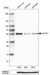
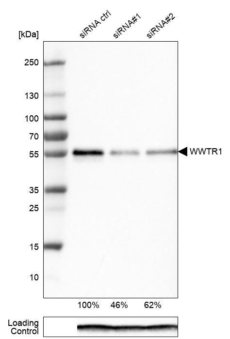
- Western blot analysis in EFO-21 cells transfected with control siRNA, target specific siRNA probe #1 and #2, using Anti-WWTR1 antibody. Remaining relative intensity is presented. Loading control: Anti-PPIB.
- Sample type
- Human
- Protocol
- Protocol
Supportive validation
- Submitted by
- Atlas Antibodies (provider)
- Main image
- Experimental details
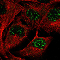
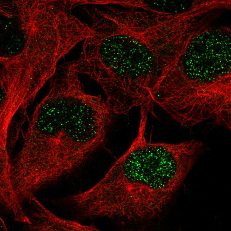
- Immunofluorescence staining in SKMEL cell line with Anti-WWTR1 monoclonal antibody, showing spotty nuclear staining in green. Microtubule probes are visualized in red (where available).
- Sample type
- Human
Supportive validation
- Submitted by
- Atlas Antibodies (provider)
- Enhanced method
- Orthogonal validation
- Main image
- Experimental details
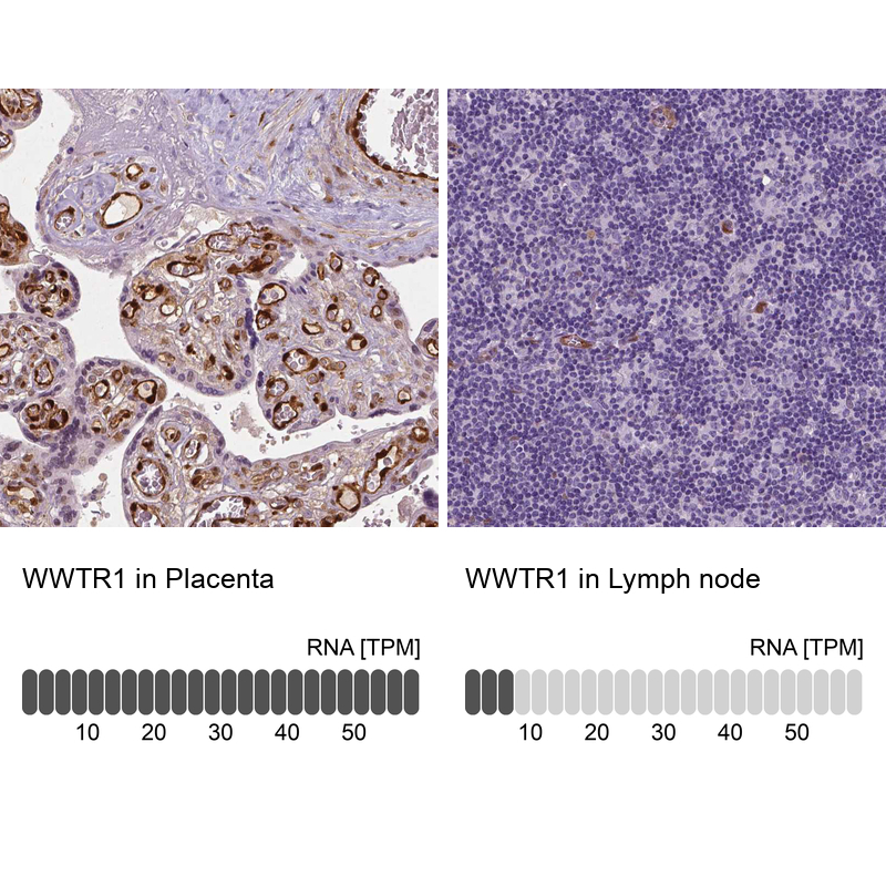
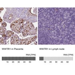
- Immunohistochemistry analysis in human placenta and lymph node tissues using AMAb90730 antibody. Corresponding WWTR1 RNA-seq data are presented for the same tissues.
- Sample type
- Human
- Protocol
- Protocol