 Explore
 Validate
 Learn
LearnPA1-46190
antibody from Invitrogen Antibodies
Targeting: WWTR1
DKFZp586I1419, TAZ
 Western blot
 Immunocytochemistry
 Immunoprecipitation Immunohistochemistry Chromatin Immunoprecipitation
Antibody data
- Antibody Data
- Antigen structure
- References [0]
- Comments [0]
- Validations
- Immunocytochemistry [2]
- Immunohistochemistry [1]
Submit
Validation data
Reference
Comment
Report error
- Product number
- PA1-46190 - Provider product page

- Provider
- Invitrogen Antibodies
- Product name
- WWTR1 Polyclonal Antibody
- Antibody type
- Polyclonal
- Antigen
- Synthetic peptide
- Description
- Suggested positive control: 293 whole cell lysate, antigen standard for WWTR1 (transient overexpression lysate), HEK 293 lysate and kidney tissue lysate, human kidney protein.
- Reactivity
- Human, Mouse, Rat
- Host
- Rabbit
- Isotype
- IgG
- Vial size
- 100 μL
- Concentration
- 1 mg/mL
- Storage
- -20°C or -80°C if preferred
No comments: Submit comment
Supportive validation
- Submitted by
- Invitrogen Antibodies (provider)
- Main image

- Experimental details
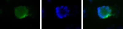
- Immunocytochemistry analysis of WWTR1 in HepG2 cells. Samples were incubated in WWTR1 polyclonal antibody (Product # PA1-46190). Nuclei (Blue) are counterstained with Hoecsht 33258.
- Submitted by
- Invitrogen Antibodies (provider)
- Main image

- Experimental details
- Immunocytochemistry analysis of WWTR1 in HepG2 cells. Samples were incubated in WWTR1 polyclonal antibody (Product # PA1-46190). Nuclei (Blue) are counterstained with Hoecsht 33258.

Supportive validation
- Submitted by
- Invitrogen Antibodies (provider)
- Main image
- Experimental details
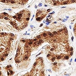
- Immunohistochemical analysis of WWTR1 in formalin fixed paraffin-embedded (FFPE) human kidney. Samples were incubated in WWTR1 polyclonal antibody (Product # PA1-46190) using a dilution of 1:500. Bond Rx autostainer (Leica Biosystems). The assay involved 20 minutes of heat induced antigen retrieval (HIER) using 10mM sodium citrate buffer (pH 6.0) and endogenous peroxidase quenching with peroxide block. The sections were incubated with primary antibody for 30 minutes and Bond Polymer Refine Detection (Leica Biosystems) with DAB was used for signal development followed by counterstaining with hematoxylin. Whole slide scanning and capturing of representative images was performed using Aperio AT2 (Leica Biosystems). Nuclear and cytoplasmic staining was observed. Staining was performed by Histowiz.
