 Explore
 Validate
 Learn
Learn Western blot
 Immunocytochemistry
 Immunohistochemistry
Antibody data
- Antibody Data
- Antigen structure
- References [0]
- Comments [0]
- Validations
- Western blot [1]
- Immunocytochemistry [3]
Submit
Validation data
Reference
Comment
Report error
- Product number
- LS-B10408 - Provider product page

- Provider
- LSBio
- Product name
- IHC-plus™ Rhodopsin / RHO Antibody (clone B630) LS-B10408
- Antibody type
- Monoclonal
- Description
- Protein G purified
- Reactivity
- Human, Mouse, Rat, Bovine, Porcine
- Host
- Mouse
- Isotype
- IgG
- Antibody clone number
- B630
- Storage
- Short term: store at 4°C. Long term: store at -20°C. Avoid freeze-thaw cycles.
No comments: Submit comment
Enhanced validation
- Submitted by
- LSBio (provider)
- Enhanced method
- Genetic validation
- Main image
- Experimental details
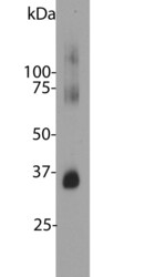
- Blot of bovine retinal extracts probed with Rhodopsin / RHO antibody. The antibody stains a band corresponding to retinal rhodopsin at about 35kDa. Bands about 70 kDa and 140 kDa are aggregated forms of rhodopsin. Note, due to the highly hydrophobic nature of rhodopsin, it important to avoid boiling samples containing this protein it in SDS-PAGE sample buffer, as this will result in even more extensive aggregation of the rhodopsin protein and appearance of more of this high molecular weight material.

Supportive validation
- Submitted by
- LSBio (provider)
- Enhanced method
- Genetic validation
- Main image
- Experimental details
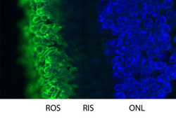
- High magnification confocal image of pig retinal section stained with Rhodopsin / RHO antibody (Green). Rhodopsin is most abundant in the rod outer segments (ROS) of retina, clearly localized in rod membranes. The rod inner segments (RIS) and rod nuclei in the outer nuclear layer (ONL) are also seen in this image. Nuclear DNA was stained with DAPI (blue).
- Submitted by
- LSBio (provider)
- Main image
- Experimental details
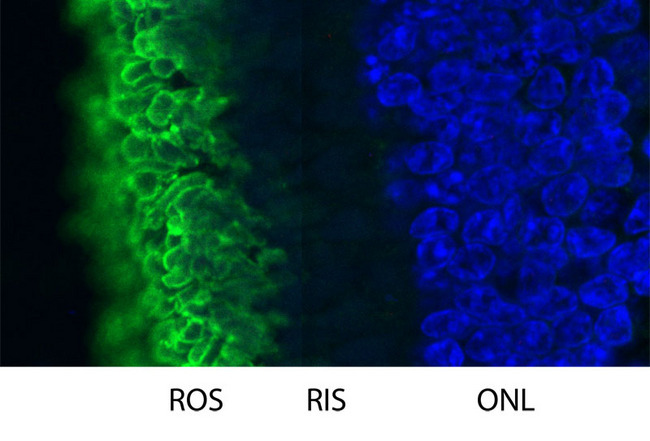
- High magnification confocal image of pig retinal section stained with Rhodopsin / RHO antibody (Green). Rhodopsin is most abundant in the rod outer segments (ROS) of retina, clearly localized in rod membranes. The rod inner segments (RIS) and rod nuclei in the outer nuclear layer (ONL) are also seen in this image. Nuclear DNA was stained with DAPI (blue).

- Submitted by
- LSBio (provider)
- Main image

- Experimental details
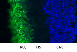
- High magnification confocal image of pig retinal section stained with Rhodopsin / RHO antibody (Green). Rhodopsin is most abundant in the rod outer segments (ROS) of retina, clearly localized in rod membranes. The rod inner segments (RIS) and rod nuclei in the outer nuclear layer (ONL) are also seen in this image. Nuclear DNA was stained with DAPI (blue).
