Explore
 Validate
 Learn
Learn Western blot
 Immunocytochemistry
 Immunohistochemistry
Antibody data
- Antibody Data
- Antigen structure
- References [0]
- Comments [0]
- Validations
- Western blot [1]
- Immunohistochemistry [1]
Submit
Validation data
Reference
Comment
Report error
- Product number
- LS-B10409 - Provider product page

- Provider
- LSBio
- Product name
- IHC-plus™ Rhodopsin / RHO Antibody (clone A531) LS-B10409
- Antibody type
- Monoclonal
- Description
- Protein G purified
- Reactivity
- Human, Mouse, Rat, Bovine, Porcine
- Host
- Mouse
- Isotype
- IgG
- Antibody clone number
- A531
- Storage
- Store at 4°C or -20°C. Avoid freeze-thaw cycles.
No comments: Submit comment
Enhanced validation
- Submitted by
- LSBio (provider)
- Enhanced method
- Genetic validation
- Main image

- Experimental details
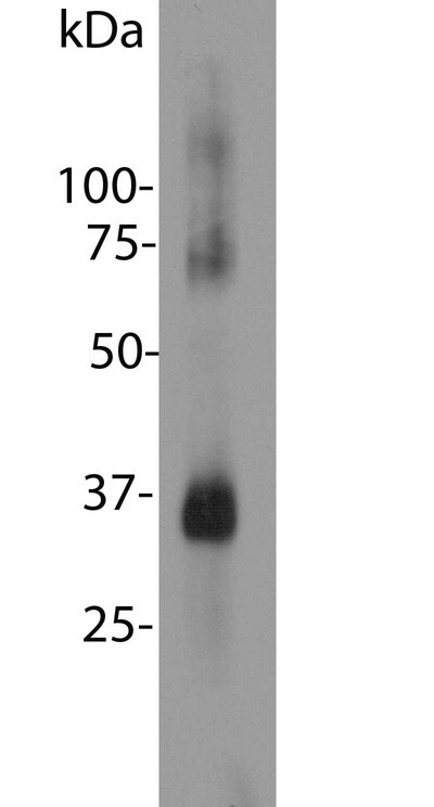
- Blot of bovine retinal extracts probed with Rhodopsin / RHO antibody. The antibody stains a band corresponding to retinal rhodopsin at about 35kDa. Bands about 70 kDa and 140 kDa are aggregated forms of rhodopsin. Note, due to the highly hydrophobic nature of rhodopsin, it is important not to boil a sample containing it in SDS-PAGE sample buffer, as this will result in more extensive aggregation of the rhodopsin protein.
Supportive validation
- Submitted by
- LSBio (provider)
- Enhanced method
- Genetic validation
- Main image
- Experimental details
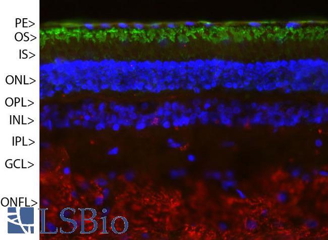
- Pig retinal section stained with Rhodopsin / RHO antibody (green) and counterstained with rabbit polyclonal antibody to neurofilament RPCA-NF-M (red) and DNA (blue). Rhodopsin is most abundant in the outer segments of retina (OS), NF-M is abundant in the optic nerve fiber layer (ONFL), but seen in processes and neurons in other regions also. Other layers are pigmented epithelium (PE), outer and inner nuclear layers (ONL, INL), outer and inner plexiform layers (OPL, IPL) and ganglion cell layer (GCL).