 Explore
 Validate
 Learn
Learn Western blot
 Immunohistochemistry
Antibody data
- Antibody Data
- Antigen structure
- References [2]
- Comments [0]
- Validations
- Western blot [1]
- Immunocytochemistry [1]
Submit
Validation data
Reference
Comment
Report error
- Product number
- ABIN1842255 - Provider product page

- Provider
- antibodies-online
- Product name
- anti-Rhodopsin (RHO) antibody
- Antibody type
- Monoclonal
- Antigen
- Other
- Reactivity
- Human, Rat, Bovine, Porcine
- Host
- Mouse
- Isotype
- IgG
- Antibody clone number
- A531
- Vial size
- 100 μL
Submitted references Photoreceptor membrane proteins, phototransduction, and retinal degenerative diseases. The Friedenwald Lecture.
Phototransduction mechanism in retinal rods and cones. The Friedenwald Lecture.
Molday RS
Investigative ophthalmology & visual science 1998 Dec;39(13):2491-513
Investigative ophthalmology & visual science 1998 Dec;39(13):2491-513
Phototransduction mechanism in retinal rods and cones. The Friedenwald Lecture.
Yau KW
Investigative ophthalmology & visual science 1994 Jan;35(1):9-32
Investigative ophthalmology & visual science 1994 Jan;35(1):9-32
No comments: Submit comment
Supportive validation
- Submitted by
- antibodies-online (provider)
- Main image
- Experimental details
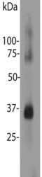
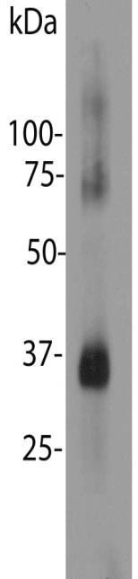
- Blot of bovine retinal extracts probed with ABIN1842255. The antibody stains a band corresponding to retinal rhodopsin at about 35 kDa. Bands about 70 kDa and 140 kDa are aggregated forms of rhodopsin. Note, due to the highly hydrophobic nature of rhodopsin, it is important not to boil a sample containing it in SDS-PAGE sample buffer, as this will result in more extensive aggregation of the rhodopsin protein.
Supportive validation
- Submitted by
- antibodies-online (provider)
- Main image
- Experimental details
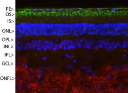
- Pig retinal section stained with ABIN1842255 (green) and counterstained with ?s rabbit polyclonal antibody to neurofilament RPCA-NF-M (red) and DNA (blue). Rhodopsin is most abundant in the outer segments of retina (OS), NF-M is abundant in the optic nerve fiber layer (ONFL), but seen in processes and neurons in other 4949 SW 41st Blvd. Suites 40 & 50 Gainesville, FL 32608 Tel: (352) 372 7022 Fax: (352) 372 7066 admin@bio.com regions also. Other layers are pigmented epithelium (PE), outer and inner nuclear layers (ONL, INL), outer and inner plexiform layers (OPL, IPL) and ganglion cell layer (GCL).
