 Explore
 Validate
 Learn
Learn Western blot
 Immunoprecipitation
 Immunohistochemistry
Antibody data
- Antibody Data
- Antigen structure
- References [33]
- Comments [0]
- Validations
- Immunohistochemistry [4]
- Other assay [5]
Submit
Validation data
Reference
Comment
Report error
- Product number
- MA1-722 - Provider product page

- Provider
- Invitrogen Antibodies
- Product name
- Rhodopsin Monoclonal Antibody (1D4)
- Antibody type
- Monoclonal
- Antigen
- Other
- Description
- MA1-722 detects rhodopsin from human, mouse and bovine retinal samples. Clone 1D4 has also been used successfully in Zebrafish. It does not recognize rhodopsin in zebrafish rods, but instead labels long double cone outer segments that express red opsin (Yin, J. et al, 2012). MA1-722 has been successfully used in Western blot, IHC (P), immunocytochemistry and immunoprecipitation procedures. By Western blot, this antibody detects an ~40 kDa protein representing rhodopsin from Sf9 cells expressing the bovine gene. Immunocytochemical staining of rhodopsin in human retinal samples results in staining of both rod and cone outer segments. The MA1-722 immunogen is bovine rhodopsin. The epitope for this antibody has been localized to the C-terminal nine amino acids of bovine rhodopsin known as the 1D4 epitope.
- Reactivity
- Human, Mouse, Rat, Bovine
- Host
- Mouse
- Isotype
- IgG
- Antibody clone number
- 1D4
- Vial size
- 100 μL
- Concentration
- 1 mg/mL
- Storage
- -20°C, Avoid Freeze/Thaw Cycles
Submitted references Effects of Epigenetic Modification of PGC-1α by a Chemical Chaperon on Mitochondria Biogenesis and Visual Function in Retinitis Pigmentosa.
Deletion of Asrgl1 Leads to Photoreceptor Degeneration in Mice.
Neuroprotective Effect of 4-Phenylbutyric Acid against Photo-Stress in the Retina.
Functional analyses of epidemic Clostridioides difficile toxin B variants reveal their divergence in utilizing receptors and inducing pathology.
SARS-CoV-2 and Three Related Coronaviruses Utilize Multiple ACE2 Orthologs and Are Potently Blocked by an Improved ACE2-Ig.
Genome-Wide CRISPR Screen Identifies Semaphorin 6A and 6B as Receptors for Paeniclostridium sordellii Toxin TcsL.
Deletion of the Impg2 gene causes the degeneration of rod and cone cells in mice.
Role of Translational Attenuation in Inherited Retinal Degeneration.
Metipranolol promotes structure and function of retinal photoreceptors in the rd10 mouse model of human retinitis pigmentosa.
Cryo-EM structure of human rhodopsin bound to an inhibitory G protein.
Whole-exome sequencing revealed HKDC1 as a candidate gene associated with autosomal-recessive retinitis pigmentosa.
A G Protein-Coupled Receptor Dimerization Interface in Human Cone Opsins.
Frizzled proteins are colonic epithelial receptors for C. difficile toxin B.
1D4: a versatile epitope tag for the purification and characterization of expressed membrane and soluble proteins.
Peripherin-2 couples rhodopsin to the CNG channel in outer segments of rod photoreceptors.
Mosaic synaptopathy and functional defects in Cav1.4 heterozygous mice and human carriers of CSNB2.
Human melanopsin forms a pigment maximally sensitive to blue light (λmax ≈ 479 nm) supporting activation of G(q/11) and G(i/o) signalling cascades.
Reproducible and sustained regulation of Gαs signalling using a metazoan opsin as an optogenetic tool.
In vitro assays of rod and cone opsin activity: retinoid analogs as agonists and inverse agonists.
Assays for inverse agonists in the visual system.
TSPAN12 regulates retinal vascular development by promoting Norrin- but not Wnt-induced FZD4/beta-catenin signaling.
High levels of retinal membrane docosahexaenoic acid increase susceptibility to stress-induced degeneration.
A neural-specific F-box protein Fbs1 functions as a chaperone suppressing glycoprotein aggregation.
Delayed loss of cone and remaining rod photoreceptor cells due to impairment of choroidal circulation after acute light exposure in rats.
Alleviation of constant-light-induced photoreceptor degeneration by adaptation of adult albino rat to bright cyclic light.
RPGRIP1s with distinct neuronal localization and biochemical properties associate selectively with RanBP2 in amacrine neurons.
A functional rhodopsin-green fluorescent protein fusion protein localizes correctly in transgenic Xenopus laevis retinal rods and is expressed in a time-dependent pattern.
Disease sequence from mutant rhodopsin allele to rod and cone photoreceptor degeneration in man.
Disease sequence from mutant rhodopsin allele to rod and cone photoreceptor degeneration in man.
Rpe65 is necessary for production of 11-cis-vitamin A in the retinal visual cycle.
Point mutations in bovine opsin can be classified in four groups with respect to their effect on the biosynthetic pathway of opsin.
Antigen-antibody interaction. Synthetic peptides define linear antigenic determinants recognized by monoclonal antibodies directed to the cytoplasmic carboxyl terminus of rhodopsin.
Monoclonal antibodies to rhodopsin: characterization, cross-reactivity, and application as structural probes.
Ozawa Y, Toda E, Homma K, Osada H, Nagai N, Tsubota K, Okano H
Cells 2022 Apr 29;11(9)
Cells 2022 Apr 29;11(9)
Deletion of Asrgl1 Leads to Photoreceptor Degeneration in Mice.
Zhou Y, Tian W, Jiang X, Yang H, Jiang Z, Li X, Jiang D, Sun K, Yang Y, Liu W, Zhu X
Frontiers in cell and developmental biology 2021;9:783547
Frontiers in cell and developmental biology 2021;9:783547
Neuroprotective Effect of 4-Phenylbutyric Acid against Photo-Stress in the Retina.
Guzmán Mendoza NA, Homma K, Osada H, Toda E, Ban N, Nagai N, Negishi K, Tsubota K, Ozawa Y
Antioxidants (Basel, Switzerland) 2021 Jul 20;10(7)
Antioxidants (Basel, Switzerland) 2021 Jul 20;10(7)
Functional analyses of epidemic Clostridioides difficile toxin B variants reveal their divergence in utilizing receptors and inducing pathology.
Pan Z, Zhang Y, Luo J, Li D, Zhou Y, He L, Yang Q, Dong M, Tao L
PLoS pathogens 2021 Jan;17(1):e1009197
PLoS pathogens 2021 Jan;17(1):e1009197
SARS-CoV-2 and Three Related Coronaviruses Utilize Multiple ACE2 Orthologs and Are Potently Blocked by an Improved ACE2-Ig.
Li Y, Wang H, Tang X, Fang S, Ma D, Du C, Wang Y, Pan H, Yao W, Zhang R, Zou X, Zheng J, Xu L, Farzan M, Zhong G
Journal of virology 2020 Oct 27;94(22)
Journal of virology 2020 Oct 27;94(22)
Genome-Wide CRISPR Screen Identifies Semaphorin 6A and 6B as Receptors for Paeniclostridium sordellii Toxin TcsL.
Tian S, Liu Y, Wu H, Liu H, Zeng J, Choi MY, Chen H, Gerhard R, Dong M
Cell host & microbe 2020 May 13;27(5):782-792.e7
Cell host & microbe 2020 May 13;27(5):782-792.e7
Deletion of the Impg2 gene causes the degeneration of rod and cone cells in mice.
Xu H, Qu C, Gan L, Sun K, Tan J, Liu X, Jiang Z, Tian W, Liu W, Zhang S, Yang Y, Jiang L, Zhu X, Zhang L
Human molecular genetics 2020 Jun 27;29(10):1624-1634
Human molecular genetics 2020 Jun 27;29(10):1624-1634
Role of Translational Attenuation in Inherited Retinal Degeneration.
Starr CR, Nyankerh CNA, Qi X, Hu Y, Gorbatyuk OS, Sonenberg N, Boulton ME, Gorbatyuk MS
Investigative ophthalmology & visual science 2019 Nov 1;60(14):4849-4857
Investigative ophthalmology & visual science 2019 Nov 1;60(14):4849-4857
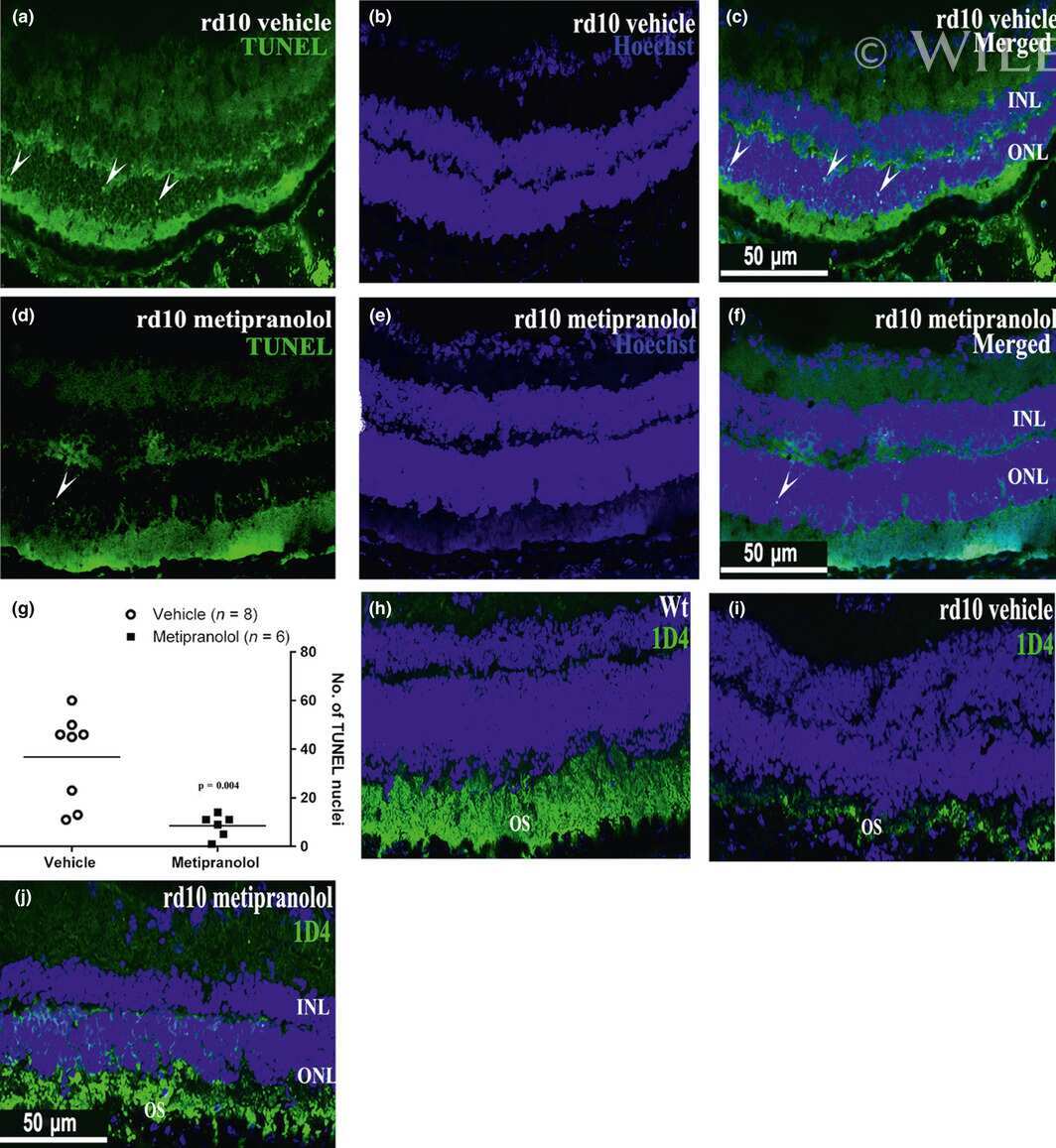
Metipranolol promotes structure and function of retinal photoreceptors in the rd10 mouse model of human retinitis pigmentosa.
Kanan Y, Khan M, Lorenc VE, Long D, Chadha R, Sciamanna J, Green K, Campochiaro PA
Journal of neurochemistry 2019 Jan;148(2):307-318
Journal of neurochemistry 2019 Jan;148(2):307-318
Cryo-EM structure of human rhodopsin bound to an inhibitory G protein.
Kang Y, Kuybeda O, de Waal PW, Mukherjee S, Van Eps N, Dutka P, Zhou XE, Bartesaghi A, Erramilli S, Morizumi T, Gu X, Yin Y, Liu P, Jiang Y, Meng X, Zhao G, Melcher K, Ernst OP, Kossiakoff AA, Subramaniam S, Xu HE
Nature 2018 Jun;558(7711):553-558
Nature 2018 Jun;558(7711):553-558
Whole-exome sequencing revealed HKDC1 as a candidate gene associated with autosomal-recessive retinitis pigmentosa.
Zhang L, Sun Z, Zhao P, Huang L, Xu M, Yang Y, Chen X, Lu F, Zhang X, Wang H, Zhang S, Liu W, Jiang Z, Ma S, Chen R, Zhao C, Yang Z, Sui R, Zhu X
Human molecular genetics 2018 Dec 1;27(23):4157-4168
Human molecular genetics 2018 Dec 1;27(23):4157-4168
A G Protein-Coupled Receptor Dimerization Interface in Human Cone Opsins.
Jastrzebska B, Comar WD, Kaliszewski MJ, Skinner KC, Torcasio MH, Esway AS, Jin H, Palczewski K, Smith AW
Biochemistry 2017 Jan 10;56(1):61-72
Biochemistry 2017 Jan 10;56(1):61-72
Frizzled proteins are colonic epithelial receptors for C. difficile toxin B.
Tao L, Zhang J, Meraner P, Tovaglieri A, Wu X, Gerhard R, Zhang X, Stallcup WB, Miao J, He X, Hurdle JG, Breault DT, Brass AL, Dong M
Nature 2016 Oct 20;538(7625):350-355
Nature 2016 Oct 20;538(7625):350-355
1D4: a versatile epitope tag for the purification and characterization of expressed membrane and soluble proteins.
Molday LL, Molday RS
Methods in molecular biology (Clifton, N.J.) 2014;1177:1-15
Methods in molecular biology (Clifton, N.J.) 2014;1177:1-15
Peripherin-2 couples rhodopsin to the CNG channel in outer segments of rod photoreceptors.
Becirovic E, Nguyen ON, Paparizos C, Butz ES, Stern-Schneider G, Wolfrum U, Hauck SM, Ueffing M, Wahl-Schott C, Michalakis S, Biel M
Human molecular genetics 2014 Nov 15;23(22):5989-97
Human molecular genetics 2014 Nov 15;23(22):5989-97
Mosaic synaptopathy and functional defects in Cav1.4 heterozygous mice and human carriers of CSNB2.
Michalakis S, Shaltiel L, Sothilingam V, Koch S, Schludi V, Krause S, Zeitz C, Audo I, Lancelot ME, Hamel C, Meunier I, Preising MN, Friedburg C, Lorenz B, Zabouri N, Haverkamp S, Garcia Garrido M, Tanimoto N, Seeliger MW, Biel M, Wahl-Schott CA
Human molecular genetics 2014 Mar 15;23(6):1538-50
Human molecular genetics 2014 Mar 15;23(6):1538-50
Human melanopsin forms a pigment maximally sensitive to blue light (λmax ≈ 479 nm) supporting activation of G(q/11) and G(i/o) signalling cascades.
Bailes HJ, Lucas RJ
Proceedings. Biological sciences 2013 May 22;280(1759):20122987
Proceedings. Biological sciences 2013 May 22;280(1759):20122987
Reproducible and sustained regulation of Gαs signalling using a metazoan opsin as an optogenetic tool.
Bailes HJ, Zhuang LY, Lucas RJ
PloS one 2012;7(1):e30774
PloS one 2012;7(1):e30774
In vitro assays of rod and cone opsin activity: retinoid analogs as agonists and inverse agonists.
Kono M, Crouch RK
Methods in molecular biology (Clifton, N.J.) 2010;652:85-94
Methods in molecular biology (Clifton, N.J.) 2010;652:85-94
Assays for inverse agonists in the visual system.
Kono M
Methods in enzymology 2010;485:213-24
Methods in enzymology 2010;485:213-24
TSPAN12 regulates retinal vascular development by promoting Norrin- but not Wnt-induced FZD4/beta-catenin signaling.
Junge HJ, Yang S, Burton JB, Paes K, Shu X, French DM, Costa M, Rice DS, Ye W
Cell 2009 Oct 16;139(2):299-311
Cell 2009 Oct 16;139(2):299-311
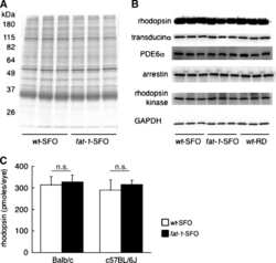
High levels of retinal membrane docosahexaenoic acid increase susceptibility to stress-induced degeneration.
Tanito M, Brush RS, Elliott MH, Wicker LD, Henry KR, Anderson RE
Journal of lipid research 2009 May;50(5):807-19
Journal of lipid research 2009 May;50(5):807-19
A neural-specific F-box protein Fbs1 functions as a chaperone suppressing glycoprotein aggregation.
Yoshida Y, Murakami A, Iwai K, Tanaka K
The Journal of biological chemistry 2007 Mar 9;282(10):7137-44
The Journal of biological chemistry 2007 Mar 9;282(10):7137-44
Delayed loss of cone and remaining rod photoreceptor cells due to impairment of choroidal circulation after acute light exposure in rats.
Tanito M, Kaidzu S, Anderson RE
Investigative ophthalmology & visual science 2007 Apr;48(4):1864-72
Investigative ophthalmology & visual science 2007 Apr;48(4):1864-72
Alleviation of constant-light-induced photoreceptor degeneration by adaptation of adult albino rat to bright cyclic light.
Li F, Cao W, Anderson RE
Investigative ophthalmology & visual science 2003 Nov;44(11):4968-75
Investigative ophthalmology & visual science 2003 Nov;44(11):4968-75
RPGRIP1s with distinct neuronal localization and biochemical properties associate selectively with RanBP2 in amacrine neurons.
Castagnet P, Mavlyutov T, Cai Y, Zhong F, Ferreira P
Human molecular genetics 2003 Aug 1;12(15):1847-63
Human molecular genetics 2003 Aug 1;12(15):1847-63
A functional rhodopsin-green fluorescent protein fusion protein localizes correctly in transgenic Xenopus laevis retinal rods and is expressed in a time-dependent pattern.
Moritz OL, Tam BM, Papermaster DS, Nakayama T
The Journal of biological chemistry 2001 Jul 27;276(30):28242-51
The Journal of biological chemistry 2001 Jul 27;276(30):28242-51
Disease sequence from mutant rhodopsin allele to rod and cone photoreceptor degeneration in man.
Cideciyan AV, Hood DC, Huang Y, Banin E, Li ZY, Stone EM, Milam AH, Jacobson SG
Proceedings of the National Academy of Sciences of the United States of America 1998 Jun 9;95(12):7103-8
Proceedings of the National Academy of Sciences of the United States of America 1998 Jun 9;95(12):7103-8
Rpe65 is necessary for production of 11-cis-vitamin A in the retinal visual cycle.
Redmond TM, Yu S, Lee E, Bok D, Hamasaki D, Chen N, Goletz P, Ma JX, Crouch RK, Pfeifer K
Nature genetics 1998 Dec;20(4):344-51
Nature genetics 1998 Dec;20(4):344-51
Point mutations in bovine opsin can be classified in four groups with respect to their effect on the biosynthetic pathway of opsin.
DeCaluwé GL, DeGrip WJ
The Biochemical journal 1996 Dec 15;320 ( Pt 3):807-15
The Biochemical journal 1996 Dec 15;320 ( Pt 3):807-15
Antigen-antibody interaction. Synthetic peptides define linear antigenic determinants recognized by monoclonal antibodies directed to the cytoplasmic carboxyl terminus of rhodopsin.
Hodges RS, Heaton RJ, Parker JM, Molday L, Molday RS
The Journal of biological chemistry 1988 Aug 25;263(24):11768-75
The Journal of biological chemistry 1988 Aug 25;263(24):11768-75
Monoclonal antibodies to rhodopsin: characterization, cross-reactivity, and application as structural probes.
Molday RS, MacKenzie D
Biochemistry 1983 Feb 1;22(3):653-60
Biochemistry 1983 Feb 1;22(3):653-60
No comments: Submit comment
Supportive validation
- Submitted by
- Invitrogen Antibodies (provider)
- Main image
- Experimental details
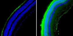
- Immunofluorescent analysis of Rhodopsin (green) showing staining in mouse retinal tissue (right) compared to a negative control without primary antibody (left). Formalin-fixed tissue was permeabilized with 0.1% Triton X-100 in TBS for 5-10 minutes and blocked with 3% BSA-PBS for 30 minutes at room temperature. Tissue was probed with a Rhodopsin monoclonal antibody (Product # MA1-722) in 3% BSA-PBS at a dilution of 1:50 and incubated overnight at 4ºC in a humidified chamber. Tissue was washed with PBST and incubated with a DyLight-488 conjugated secondary antibody in PBS at room temperature in the dark. Nuclei were stained with DAPI (blue) and images were taken at a magnification of 20x.
- Submitted by
- Invitrogen Antibodies (provider)
- Main image

- Experimental details
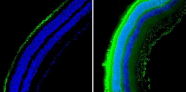
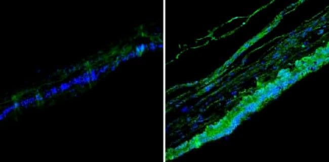
- Immunofluorescent analysis of Rhodopsin (green) showing staining in human retinal tissue (right) compared to a negative control without primary antibody (left). Formalin-fixed tissue was permeabilized with 0.1% Triton X-100 in TBS for 5-10 minutes and blocked with 3% BSA-PBS for 30 minutes at room temperature. Tissue was probed with a Rhodopsin monoclonal antibody (Product # MA1-722) in 3% BSA-PBS at a dilution of 1:50 and incubated overnight at 4ºC in a humidified chamber. Tissue was washed with PBST and incubated with a DyLight-488 conjugated secondary antibody in PBS at room temperature in the dark. Nuclei were stained with DAPI (blue) and images were taken at a magnification of 20x.
- Submitted by
- Invitrogen Antibodies (provider)
- Main image
- Experimental details
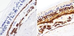
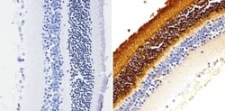
- Immunohistochemistry analysis of Rhodopsin showing staining in the nucleus of paraffin-embedded human retinal tissue (right) compared to a negative control without primary antibody (left). To expose target proteins, antigen retrieval was performed using 10mM sodium citrate (pH 6.0), microwaved for 8-15 min. Following antigen retrieval, tissues were blocked in 3% H2O2-methanol for 15 min at room temperature, washed with ddH2O and PBS, and then probed with a Rhodopsin monoclonal antibody (Product # MA1-722) diluted in 3% BSA-PBS at a dilution of 1:200 overnight at 4°C in a humidified chamber. Tissues were washed extensively in PBST and detection was performed using an HRP-conjugated secondary antibody followed by colorimetric detection using a DAB kit. Tissues were counterstained with hematoxylin and dehydrated with ethanol and xylene to prep for mounting.

- Submitted by
- Invitrogen Antibodies (provider)
- Main image
- Experimental details
- Immunohistochemistry analysis of Rhodopsin showing staining in the nucleus of paraffin-embedded mouse retinal tissue (right) compared to a negative control without primary antibody (left). To expose target proteins, antigen retrieval was performed using 10mM sodium citrate (pH 6.0), microwaved for 8-15 min. Following antigen retrieval, tissues were blocked in 3% H2O2-methanol for 15 min at room temperature, washed with ddH2O and PBS, and then probed with a Rhodopsin monoclonal antibody (Product # MA1-722) diluted in 3% BSA-PBS at a dilution of 1:1000 overnight at 4°C in a humidified chamber. Tissues were washed extensively in PBST and detection was performed using an HRP-conjugated secondary antibody followed by colorimetric detection using a DAB kit. Tissues were counterstained with hematoxylin and dehydrated with ethanol and xylene to prep for mounting.

Supportive validation
- Submitted by
- Invitrogen Antibodies (provider)
- Main image
- Experimental details
- NULL
- Submitted by
- Invitrogen Antibodies (provider)
- Main image
- Experimental details
- NULL

- Submitted by
- Invitrogen Antibodies (provider)
- Main image
- Experimental details
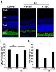
- Figure 2 4-PBA suppressed light-induced photoreceptor histological changes and degeneration. ( A ) Representative images of immunohistochemistry for rhodopsin (green) counterstained with 4',6-diamidino-2-phenylindole (DAPI) (blue) 4 days after light exposure with or without 4-PBA treatment. Reduction in ONL thickness ( B ) and OS length ( C ) were suppressed by 4-PBA. LE, light exposure; Veh, vehicle; GCL, ganglion cell layer; INL, inner nuclear layer; ONL, outer nuclear layer; OS, outer segment. Data are presented as mean +- standard deviation. n = 8 in each group. Scale bar, 25 mum. * p < 0.05, ** p < 0.01, *** p < 0.001. ANOVA with Tukey post-hoc tests.

- Submitted by
- Invitrogen Antibodies (provider)
- Main image

- Experimental details
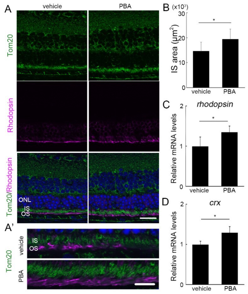
- Protective effect of PBA in the photoreceptors of P23H RP models detected before photoreceptor loss ( A , A' ) Immunohistochemistry for Tom20 (green) and rhodopsin (pink) in the retina of P23H RP models at the age of 4 weeks with or without PBA treatment. Nuclei were counterstained with DAPI (blue). ( A ') Magnified images of IS and OS. IS area ( B ), and mRNA levels of rhodopsin ( C ) and crx ( D ) in the retina were already different with or without PBA treatment. Data are shown as mean +- standard deviation. Respective n for P23H RP models treated with vehicle and PBA, ( B ) 4 and 5; ( C ) 5 and 4; ( D ) 10 and 10. ONL, outer nuclear layer; IS, inner segments; OS, outer segments. * p < 0.05. Scale bar, ( A ) 20 mum; ( B ) 10 mum.
- Submitted by
- Invitrogen Antibodies (provider)
- Main image
- Experimental details
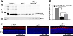
- Figure 5 Detection of rhodopsin levels in the retinas of all three mouse groups. (A) Western blot analysis was performed at P15 and the retinal protein extracts were run in SDS polyacrylamide gel and transferred to membrane. Images of membrane probed with anti-rhodopsin (upper) and anti-actin (lower) antibodies are shown. (B) The level of rhodopsin was calculated in all three groups. (C) Rhodopsin staining of retinal sections from the three groups at P15. Red, rhodopsin; blue, DAPI. Error bars: SEM. Statistical significance denoted by *P < 0.05, **P < 0.01, ****P < 0.001.
