Explore
 Validate
 Learn
Learn Western blot
 Other assay
Antibody data
- Antibody Data
- Antigen structure
- References [1]
- Comments [0]
- Validations
- Other assay [4]
Submit
Validation data
Reference
Comment
Report error
- Product number
- PA5-40508 - Provider product page

- Provider
- Invitrogen Antibodies
- Product name
- SOX30 Polyclonal Antibody
- Antibody type
- Polyclonal
- Antigen
- Synthetic peptide
- Description
- Peptide sequence: PAANNAEISV QLGLEWNKLS EEQKKPYYDE AQKIKEKHRE EFPGWVYQPR Sequence homology: Cow: 100%; Dog: 100%; Guinea Pig: 100%; Horse: 100%; Human: 100%; Mouse: 100%; Rabbit: 100%; Rat: 100%; Yeast: 100%
- Reactivity
- Human
- Host
- Rabbit
- Isotype
- IgG
- Vial size
- 100 μL
- Concentration
- 0.5 mg/mL
- Storage
- -20°C, Avoid Freeze/Thaw Cycles
Submitted references SOX30, a target gene of miR-653-5p, represses the proliferation and invasion of prostate cancer cells through inhibition of Wnt/β-catenin signaling.
Fu Q, Sun Z, Yang F, Mao T, Gao Y, Wang H
Cellular & molecular biology letters 2019;24:71
Cellular & molecular biology letters 2019;24:71
No comments: Submit comment
Supportive validation
- Submitted by
- Invitrogen Antibodies (provider)
- Main image
- Experimental details
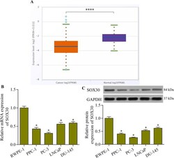
- Fig. 1 SOX30 expression was lower in prostate cancer cells than in normal prostate epithelial cells. The prostate cancer cell lines for the experiments were PPC-1, PC-3, LNCaP and DU-145. The normal prostate epithelial cell line RWPE-1 served as the control. a SOX30 expression in prostate cancer ( n = 499) and normal ( n = 52) samples was determined with the starBase Pan-Cancer Analysis Platform. **** p < 0.0001. b Relative SOX30 mRNA expression in prostate cancer cell lines was examined using quantitative real-time PCR ( n = 5, * p < 0.05 versus RWPE-1). c SOX30 protein expression in cancer cell lines was detected using western blotting ( n = 5, * p < 0.05 versus RWPE-1)

- Submitted by
- Invitrogen Antibodies (provider)
- Main image
- Experimental details
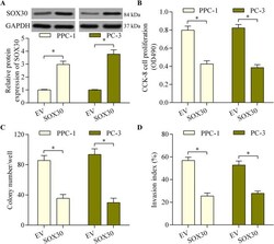
- Fig. 2 SOX30 overexpression repressed prostate cancer cell proliferation and invasion. a PPC-1 and PC-3 cells were transfected with pcDNA3.1/SOX30 vector or empty vector (EV) for 48 h and SOX30 protein expression was examined via western blotting. b The effect of SOX30 overexpression on prostate cancer cell proliferation was determined with the CCK-8 assay after transection of cells with the pcDNA3.1/SOX30 vector for 48 h. c The effect of SOX30 overexpression on prostate cancer cell colony-forming ability was assessed with a colony formation assay. PPC-1 and PC-3 cells were transfected with pcDNA3.1/SOX30 vector for 48 h and then cultured for 14 days to form colonies. d The effect of SOX30 overexpression on prostate cancer cell invasive potential was evaluated using a transwell Matrigel invasion assay. PPC-1 and PC-3 cells were transfected with pcDNA3.1/SOX30 vector for 48 h. The transwell Matrigel invasion assay ran for 24 h. ( n = 5, * p < 0.05)

- Submitted by
- Invitrogen Antibodies (provider)
- Main image
- Experimental details
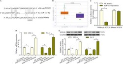
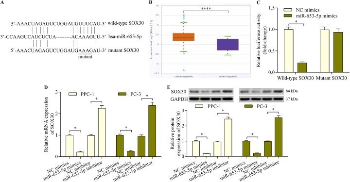
- Fig. 3 SOX30 is an miR-653-5p target gene. a Sequence alignment of the miR-653-5p-binding site within the SOX30 3'-UTR. b MiR-653-5p expression in prostate cancer ( n = 495) and normal ( n = 52) samples was determined with the starBase Pan-Cancer Analysis Platform. PPC-1, PC-3, LNCaP and DU-145 were the prostate cancer cell lines. The normal prostate epithelial cell line RWPE-1 served as the control. **** p < 0.0001. c The interaction between miR-653-5p and SOX30 3'-UTR was assessed with a dual-luciferase reporter assay using 293 T cells. SOX30 3'-UTR reporter plasmids and miR-653-5p mimics or NC mimics were co-transfected into 293 T cells and incubated for 48 h before determination of luciferase activity. n = 5, * p < 0.05. d and e PPC-1 and PC-3 cells were transfected with miR-653-5p mimics, inhibitor or NC mimics/inhibitor for 48 h. SOX30 mRNA ( d ) and protein ( e ) expressions were respectively determined using quantitative real-time PCR and western blot analysis ( n = 5, * p < 0.05)
- Submitted by
- Invitrogen Antibodies (provider)
- Main image
- Experimental details
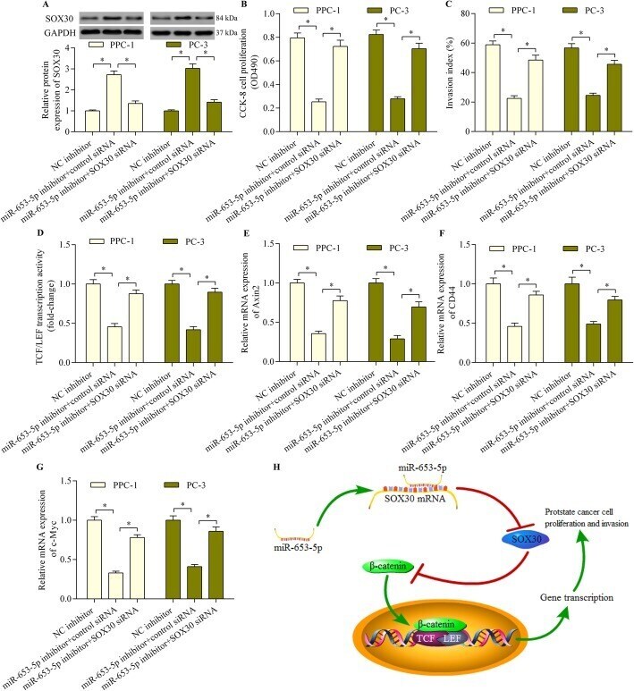
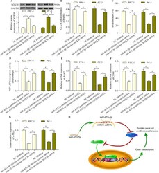
- Fig. 6 SOX30 silencing reversed the miR-653-3p inhibition-mediated anti-tumor effect in prostate cancer cells. a PPC-1 and PC-3 cells were co-transfected with miR-653-5p inhibitor and SOX30 siRNA for 48 h. SOX30 protein expression was determined using western blotting. b Cell proliferation was examined with the CCK-8 assay after co-transfection with miR-653-5p inhibitor and SOX30 siRNA for 48 h. c PPC-1 and PC-3 cells were co-transfected with miR-653-5p inhibitor and SOX30 siRNA for 48 h. A transwell Matrigel invasion assay was run for 24 h to determine cell invasion. d Wnt/beta-catenin signaling was assessed with a TCF/LEF luciferase reporter assay. PPC-1 and PC-3 cells were co-transfected with TCF/LEF luciferase reporter vector, miR-653-5p inhibitor and SOX30 siRNA and incubated for 48 h before determination of luciferase activity. e , f and g PPC-1 and PC-3 cells were co-transfected with miR-653-5p inhibitor and SOX30 siRNA for 48 h, and relative mRNA expression levels of Axin2 ( e ), CD44 ( f ), and c-Myc ( g ) were determined using quantitative real-time PCR ( n = 5, * p < 0.05). h A graphical model of the miR-653-5p-SOX30-Wnt/beta-catenin axis in regulating prostate cancer cell proliferation and invasion