Explore
 Validate
 Learn
Learn Western blot
 ELISA
ELISAAntibody data
- Antibody Data
- Antigen structure
- References [3]
- Comments [0]
- Validations
- Western blot [1]
Submit
Validation data
Reference
Comment
Report error
- Product number
- A01233-2 - Provider product page

- Provider
- Boster Biological Technology
- Product name
- Anti-TSG101 Antibody Picoband™
- Antibody type
- Polyclonal
- Description
- Rabbit IgG polyclonal antibody for TSG101 detection. Tested with WB, FCM, Direct ELISA in Human;Mouse;Rat.
- Reactivity
- Human, Mouse, Rat
- Host
- Rabbit
- Vial size
- 100μg/vial
- Concentration
- Add 0.2ml of distilled water will yield a concentration of 500ug/ml.
- Storage
- At -20°C for one year. After reconstitution, at 4°C for one month. It can also be aliquoted and stored frozen at -20°C for a longer time. Avoid repeated freezing and thawing.
- Handling
- Add 0.2ml of distilled water will yield a concentration of 500ug/ml.
Submitted references Serum exosomes derived from spontaneously hypertensive rats induce cardiac hypertrophy in vitro and in vivo by increasing autocrine release of angiotensin II in cardiomyocytes.
Exosomes from artesunate-treated bone marrow-derived mesenchymal stem cells transferring SNHG7 to promote osteogenesis via TAF15-RUNX2 pathway.
Exosome-mediated siRNA delivery to suppress postoperative breast cancer metastasis.
Yu J, Tang Y, Wang Y, Zhou M, Li Y, Hong J, Li C, Xu B, Guo X, Mao J
Biochemical pharmacology 2023 Apr;210:115462
Biochemical pharmacology 2023 Apr;210:115462
Exosomes from artesunate-treated bone marrow-derived mesenchymal stem cells transferring SNHG7 to promote osteogenesis via TAF15-RUNX2 pathway.
Huang MZ, Chen HY, Peng GX, Sun H, Peng HC, Li HY, Liu XH, Li Q
Regenerative medicine 2022 Nov;17(11):819-833
Regenerative medicine 2022 Nov;17(11):819-833
Exosome-mediated siRNA delivery to suppress postoperative breast cancer metastasis.
Zhao L, Gu C, Gan Y, Shao L, Chen H, Zhu H
Journal of controlled release : official journal of the Controlled Release Society 2020 Feb;318:1-15
Journal of controlled release : official journal of the Controlled Release Society 2020 Feb;318:1-15
No comments: Submit comment
Supportive validation
- Submitted by
- Boster Biological Technology (provider)
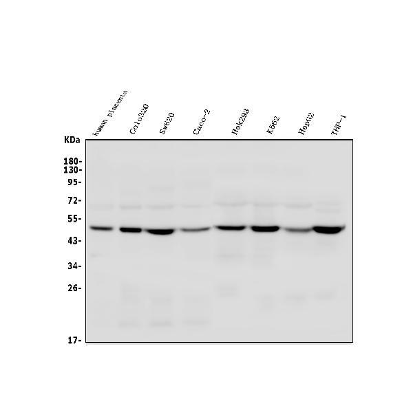
- Main image
- Experimental details
- Western blot analysis of TSG101 using anti-TSG101 antibody (A01233-2). Electrophoresis was performed on a 5-20% SDS-PAGE gel at 70V (Stacking gel) / 90V (Resolving gel) for 2-3 hours. The sample well of each lane was loaded with 50ug of sample under reducing conditions. Lane 1: human placenta tissue lysates, Lane 2: human Colo320 whole cell lysates, Lane 3: human Sw620 whole cell lysates, Lane 4: human Caco-2 whole cell lysates, Lane 5: human Hek293 whole cell lysates, Lane 6: human K562 whole cell lysates, Lane 7: human HepG2 whole cell lysates, Lane 8: human THP-1 whole cell lysates. After Electrophoresis, proteins were transferred to a Nitrocellulose membrane at 150mA for 50-90 minutes. Blocked the membrane with 5% Non-fat Milk/ TBS for 1.5 hour at RT. The membrane was incubated with rabbit anti-TSG101 antigen affinity purified polyclonal antibody (Catalog # A01233-2) at 0.25 μg/mL overnight at 4°C, then washed with TBS-0.1%Tween 3 times with 5 minutes each and probed with a goat anti-rabbit IgG-HRP secondary antibody at a dilution of 1:5000 for 1.5 hour at RT. The signal is developed using an Enhanced Chemiluminescent detection (ECL) kit (Catalog # EK1002) with Tanon 5200 system. A specific band was detected for TSG101 at approximately 44KD. The expected band size for TSG101 is at 44KD.
- Additional image