Explore
 Validate
 Learn
Learn Immunohistochemistry
Antibody data
- Antibody Data
- Antigen structure
- References [2]
- Comments [1]
- Validations
- Immunohistochemistry [1]
Submit
Validation data
Reference
Comment
Report error
- Product number
- HPA006162 - Provider product page

- Provider
- Atlas Antibodies
- Proper citation
- Atlas Antibodies Cat#HPA006162, RRID:AB_1857403
- Product name
- Anti-SP140
- Antibody type
- Polyclonal
- Description
- Polyclonal Antibody against Human SP140, Gene description: SP140 nuclear body protein, Alternative Gene Names: LYSP100-A, LYSP100-B, Validated applications: IHC, Uniprot ID: Q13342, Storage: Store at +4°C for short term storage. Long time storage is recommended at -20°C.
- Reactivity
- Human
- Host
- Rabbit
- Conjugate
- Unconjugated
- Isotype
- IgG
- Vial size
- 100 µl
- Concentration
- 0.2 mg/ml
- Storage
- Store at +4°C for short term storage. Long time storage is recommended at -20°C.
- Handling
- The antibody solution should be gently mixed before use.
Submitted references Modulation of macrophage inflammatory function through selective inhibition of the epigenetic reader protein SP140
Maintenance of macrophage transcriptional programs and intestinal homeostasis by epigenetic reader SP140
Ghiboub M, Koster J, Craggs P, Li Yim A, Shillings A, Hutchinson S, Bingham R, Gatfield K, Hageman I, Yao G, O’Keefe H, Coffin A, Patel A, Sloan L, Mitchell D, Hayhow T, Lunven L, Watson R, Blunt C, Harrison L, Bruton G, Kumar U, Hamer N, Spaull J, Zwijnenburg D, Welting O, Hakvoort T, te Velde A, van Limbergen J, Henneman P, Prinjha R, de Winther M, Harker N, Tough D, de Jonge W
BMC Biology 2022;20(1)
BMC Biology 2022;20(1)
Maintenance of macrophage transcriptional programs and intestinal homeostasis by epigenetic reader SP140
Mehta S, Cronkite D, Basavappa M, Saunders T, Adiliaghdam F, Amatullah H, Morrison S, Pagan J, Anthony R, Tonnerre P, Lauer G, Lee J, Digumarthi S, Pantano L, Ho Sui S, Ji F, Sadreyev R, Zhou C, Mullen A, Kumar V, Li Y, Wijmenga C, Xavier R, Means T, Jeffrey K
Science Immunology 2017;2(9)
Science Immunology 2017;2(9)
2011-03-28
- Submitted by
- sternsdorf
- Comment
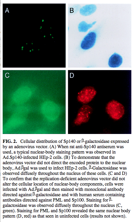
- The IF shown does not resemble the localization described for the Sp140 protein, which is typically localized in the nucleus in the form of characteristic speckles (hence the name Sp, which stands for the Speckled localization). Instead the depicted IF image resembles a staining for cytoskeletal filaments (Actin/Tubulin) and could result from nonspecific staining rather than true recognition of the Sp140antigen.As the Sp140 protein is not ubiquitously expressed, it may be necessary to use lymphoid cells for proper localization. The attached file is from the corresponding publication by Bloch et al 1999 (MCB) Sincerely, Thomas Sternsdorf Ph. D.
- Antibody rating
- 2. Poor
- Image
Supportive validation
- Submitted by
- Atlas Antibodies (provider)
- Enhanced method
- Orthogonal validation
- Main image
- Experimental details
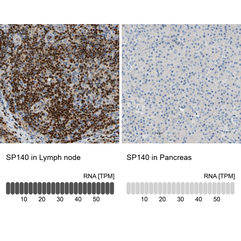
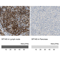
- Immunohistochemistry analysis in human lymph node and pancreas tissues using HPA006162 antibody. Corresponding SP140 RNA-seq data are presented for the same tissues.
- Sample type
- Human
- Protocol
- Protocol