Explore
 Validate
 Learn
Learn Western blot
Antibody data
- Antibody Data
- Antigen structure
- References [4]
- Comments [0]
- Validations
- Western blot [2]
- Immunocytochemistry [3]
- Immunohistochemistry [1]
Submit
Validation data
Reference
Comment
Report error
- Product number
- GTX109459 - Provider product page

- Provider
- GeneTex
- Proper citation
- GeneTex Cat#GTX109459, RRID:AB_2038129
- Product name
- TKTL1 antibody [N1C1]
- Antibody type
- Polyclonal
- Reactivity
- Human
- Host
- Rabbit
Submitted references Transketolase Regulates the Metabolic Switch to Control Breast Cancer Cell Metastasis via the α-Ketoglutarate Signaling Pathway.
Downregulation of the Werner syndrome protein induces a metabolic shift that compromises redox homeostasis and limits proliferation of cancer cells
Downregulation of the Werner syndrome protein induces a metabolic shift that compromises redox homeostasis and limits proliferation of cancer cells.
Downregulation of the Werner syndrome protein induces a metabolic shift that compromises redox homeostasis and limits proliferation of cancer cells.
Tseng CW, Kuo WH, Chan SH, Chan HL, Chang KJ, Wang LH
Cancer research 2018 Jun 1;78(11):2799-2812
Cancer research 2018 Jun 1;78(11):2799-2812
Downregulation of the Werner syndrome protein induces a metabolic shift that compromises redox homeostasis and limits proliferation of cancer cells
Li B, Iglesias-Pedraz J, Chen L, Yin F, Cadenas E, Reddy S, Comai L
Aging Cell 2014 April;13(2):367-378
Aging Cell 2014 April;13(2):367-378
Downregulation of the Werner syndrome protein induces a metabolic shift that compromises redox homeostasis and limits proliferation of cancer cells.
Li B, Iglesias-Pedraz JM, Chen LY, Yin F, Cadenas E, Reddy S, Comai L
Aging cell 2014 Apr;13(2):367-78
Aging cell 2014 Apr;13(2):367-78
Downregulation of the Werner syndrome protein induces a metabolic shift that compromises redox homeostasis and limits proliferation of cancer cells.
Li B, Iglesias-Pedraz JM, Chen LY, Yin F, Cadenas E, Reddy S, Comai L
Aging cell 2014 Apr;13(2):367-78
Aging cell 2014 Apr;13(2):367-78
No comments: Submit comment
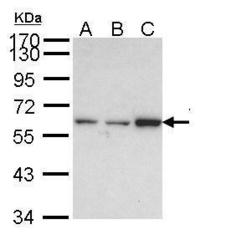
Supportive validation
- Submitted by
- GeneTex (provider)
- Main image
- Experimental details
- Sample (30 ?g of whole cell lysate) A: HeLa B: HepG2 (GTX27900) C: Molt-4 (GTX27912) 7.5% SDS PAGE GTX109459 diluted at 1:5000 The HRP-conjugated anti-rabbit IgG antibody (GTX213110-01) was used to detect the primary antibody.

- Submitted by
- GeneTex (provider)
- Main image
- Experimental details
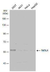
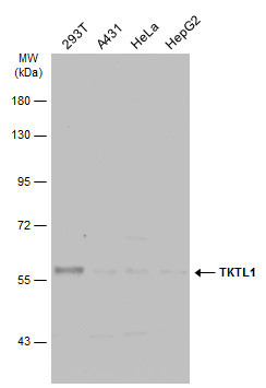
- Various whole cell extracts (30 ?g) were separated by 7.5% SDS-PAGE, and the membrane was blotted with TKTL1 antibody [N1C1] (GTX109459) diluted at 1:5000. The HRP-conjugated anti-rabbit IgG antibody (GTX213110-01) was used to detect the primary antibody.
Supportive validation
- Submitted by
- GeneTex (provider)
- Main image
- Experimental details
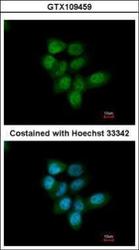
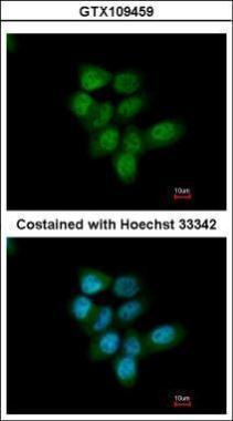
- Immunofluorescence analysis of paraformaldehyde-fixed A431, using TKTL1(GTX109459) antibody at 1:200 dilution.
- Submitted by
- GeneTex (provider)
- Main image
- Experimental details
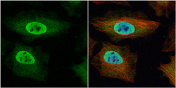
- TKTL1 antibody [N1C1] detects TKTL1 protein at cytoplasm and nucleus by immunofluorescent analysis.Sample: HeLa cells were fixed in 4% paraformaldehyde at RT for 15 min.Green: TKTL1 protein stained by TKTL1 antibody [N1C1] (GTX109459) diluted at 1:200.Red: alpha Tubulin, a cytoskeleton marker, stained by alpha Tubulin antibody [B-5-1-2] (GTX11304) diluted at 1:10000.Blue: Hoechst 33342 staining.

- Submitted by
- GeneTex (provider)
- Main image
- Experimental details
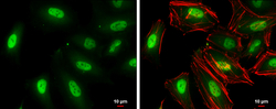
- TKTL1 antibody [N1C1] detects TKTL1 protein at cytoplasm and nucleus by immunofluorescent analysis.Sample: HeLa cells were fixed in 4% paraformaldehyde at RT for 15 min.Green: TKTL1 protein stained by TKTL1 antibody [N1C1] (GTX109459) diluted at 1:500.Red: Phalloidin, a cytoskeleton marker, diluted at 1:100.Scale bar = 10 £gm.

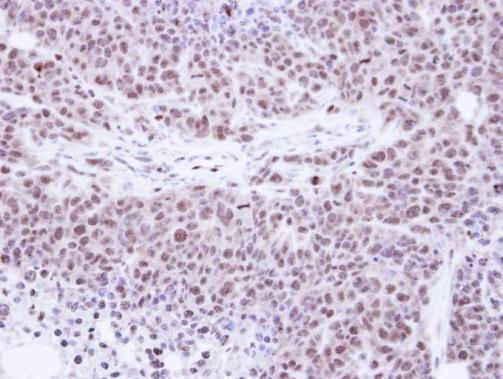
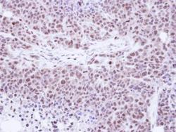
Supportive validation
- Submitted by
- GeneTex (provider)
- Main image
- Experimental details
- Immunohistochemical analysis of paraffin-embedded SAS xenograft , using Transketolase-like protein 1(GTX109459) antibody at 1:100 dilution.