Explore
 Validate
 Learn
Learn Western blot
 Immunocytochemistry
Antibody data
- Antibody Data
- Antigen structure
- References [5]
- Comments [0]
- Validations
- Immunocytochemistry [2]
- Flow cytometry [1]
- Other assay [1]
Submit
Validation data
Reference
Comment
Report error
- Product number
- 44-944G - Provider product page

- Provider
- Invitrogen Antibodies
- Product name
- Phospho-NFATC2 (Ser54) Polyclonal Antibody
- Antibody type
- Polyclonal
- Antigen
- Synthetic peptide
- Reactivity
- Human, Mouse
- Host
- Rabbit
- Isotype
- IgG
- Vial size
- 100 μL
- Storage
- -20°C
Submitted references Evidence for reprogramming of monocytes into reparative alveolar macrophages in vivo by targeting PDE4b.
Overexpression of DSCR1 prevents proliferation and predicts favorable prognosis in colorectal cancer patients.
STIM1 controls calcineurin/Akt/mTOR/NFATC2-mediated osteoclastogenesis induced by RANKL/M-CSF.
Regulation of phagocytosis and cytokine secretion by store-operated calcium entry in primary isolated murine microglia.
NK cell-activating receptors require PKC-theta for sustained signaling, transcriptional activation, and IFN-gamma secretion.
Rochford I, Joshi JC, Rayees S, Anwar M, Akhter MZ, Yalagala L, Banerjee S, Mehta D
American journal of physiology. Lung cellular and molecular physiology 2021 Oct 1;321(4):L686-L702
American journal of physiology. Lung cellular and molecular physiology 2021 Oct 1;321(4):L686-L702
Overexpression of DSCR1 prevents proliferation and predicts favorable prognosis in colorectal cancer patients.
Li WX, Zheng JJ, Zhao G, Lyu CT, Lu WQ
World journal of surgical oncology 2021 Apr 7;19(1):100
World journal of surgical oncology 2021 Apr 7;19(1):100
STIM1 controls calcineurin/Akt/mTOR/NFATC2-mediated osteoclastogenesis induced by RANKL/M-CSF.
Huang Y, Li Q, Feng Z, Zheng L
Experimental and therapeutic medicine 2020 Aug;20(2):736-747
Experimental and therapeutic medicine 2020 Aug;20(2):736-747
Regulation of phagocytosis and cytokine secretion by store-operated calcium entry in primary isolated murine microglia.
Heo DK, Lim HM, Nam JH, Lee MG, Kim JY
Cellular signalling 2015 Jan;27(1):177-86
Cellular signalling 2015 Jan;27(1):177-86
NK cell-activating receptors require PKC-theta for sustained signaling, transcriptional activation, and IFN-gamma secretion.
Tassi I, Cella M, Presti R, Colucci A, Gilfillan S, Littman DR, Colonna M
Blood 2008 Nov 15;112(10):4109-16
Blood 2008 Nov 15;112(10):4109-16
No comments: Submit comment
Supportive validation
- Submitted by
- Invitrogen Antibodies (provider)
- Main image

- Experimental details
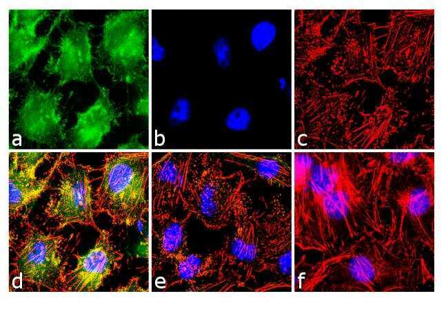
- Immunofluorescence analysis of Phospho-NFAT1 (Ser54) was done on 70% confluent log phase HeLa cells treated with 200nM of PMA for 20 minutes. The cells were fixed with 4% paraformaldehyde for 10 minutes, permeabilized with 0.1% Triton™ X-100 for 10 minutes, and blocked with 1% BSA for 1 hour at room temperature. The cells were labeled with phospho-NFAT1 (Ser54) Rabbit Polyclonal Antibody (Product # 44-944G) at 1:250 dilution in 0.1% BSA and incubated for 3 hours at room temperature and then labeled with Goat anti-Rabbit IgG (H+L) Superclonal™ Secondary Antibody, Alexa Fluor® 488 conjugate (Product # A27034) at a dilution of 1:2000 for 45 minutes at room temperature (Panel a: green). Nuclei (Panel b: blue) were stained with SlowFade® Gold Antifade Mountant with DAPI (Product # S36938). F-actin (Panel c: red) was stained with Alexa Fluor® 555 Rhodamine Phalloidin (Product # R415, 1:300). Panel d is a merged image showing cytoplasmic and nuclear localization. Panel e is untreated cells with no signal. Panel f is no primary antibody control. The images were captured at 60X magnification.
- Submitted by
- Invitrogen Antibodies (provider)
- Main image

- Experimental details
- Immunofluorescence analysis of Phospho-NFAT1 (Ser54) was done on 70% confluent log phase HeLa cells treated with 200nM of PMA for 20 minutes. The cells were fixed with 4% paraformaldehyde for 10 minutes, permeabilized with 0.1% Triton™ X-100 for 10 minutes, and blocked with 1% BSA for 1 hour at room temperature. The cells were labeled with phospho-NFAT1 (Ser54) Rabbit Polyclonal Antibody (Product # 44-944G) at 1:250 dilution in 0.1% BSA and incubated for 3 hours at room temperature and then labeled with Goat anti-Rabbit IgG (Heavy Chain) Superclonal™ Secondary Antibody, Alexa Fluor® 488 conjugate (Product # A27034) at a dilution of 1:2000 for 45 minutes at room temperature (Panel a: green). Nuclei (Panel b: blue) were stained with SlowFade® Gold Antifade Mountant with DAPI (Product # S36938). F-actin (Panel c: red) was stained with Alexa Fluor® 555 Rhodamine Phalloidin (Product # R415, 1:300). Panel d is a merged image showing cytoplasmic and nuclear localization. Panel e is untreated cells with no signal. Panel f is no primary antibody control. The images were captured at 60X magnification.

Supportive validation
- Submitted by
- Invitrogen Antibodies (provider)
- Main image
- Experimental details
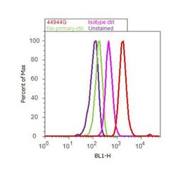
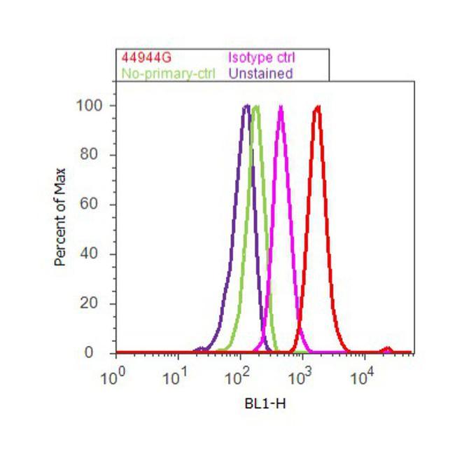
- Flow cytometry analysis of NFAT1 [pSer54] was done on HeLa cells treated with PMA (200nM, 20 minutes). Cells were fixed with 70% ethanol for 10 minutes, permeabilized with 0.25% Triton™ X-100 for 20 minutes, and blocked with 5% BSA for 30 minutes at room temperature. Cells were labeled with NFAT1 [pSer54] Rabbit Polyclonal Antibody (44944G, red histogram) or with rabbit isotype control (pink histogram) at 3-5 ug/million cells in 2.5% BSA. After incubation at room temperature for 2 hours, the cells were labeled with Alexa Fluor® 488 Goat Anti-Rabbit Secondary Antibody (A11008) at a dilution of 1:400 for 30 minutes at room temperature. The representative 10,000 cells were acquired and analyzed for each sample using an Attune® Acoustic Focusing Cytometer. The purple histogram represents unstained control cells and the green histogram represents no-primary-antibody control.
Supportive validation
- Submitted by
- Invitrogen Antibodies (provider)
- Main image
- Experimental details
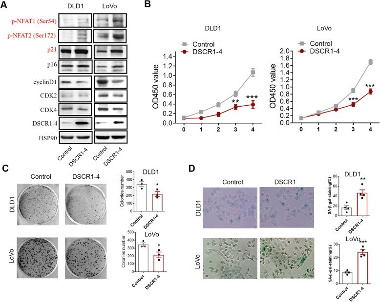
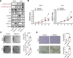
- Fig. 3 Overexpression of DSCR1-4 showed tumor suppressive function in CRC cells in vitro. a Western blot showed that overexpression of DSCR1-4 increased p21, p16, and hyperphosphorylation of NFAT1 and NFAT2, while decreased cyclin D1, CDK2, and CDK4. b CCK8 assays showed that overexpression of DSCR1-4 inhibited cell proliferation of CRC cells. Data are mean +- SEM of 5 replicates. c Overexpression of DSCR1-4 inhibited cell colony formation of CRC cells. Data are mean +- SEM of 3 replicates. d Overexpression of DSCR1-4 induced senescence of CRC cells by SA-beta-gal staining. Two-tailed Student t test, * p < 0.05, ** p < 0.01, *** p < 0.001