Explore
 Validate
 Learn
Learn Western blot
 Immunohistochemistry
Antibody data
- Antibody Data
- Antigen structure
- References [3]
- Comments [0]
- Validations
- Immunohistochemistry [2]
- Other assay [2]
Submit
Validation data
Reference
Comment
Report error
- Product number
- PA1-108 - Provider product page

- Provider
- Invitrogen Antibodies
- Product name
- PAX4 Polyclonal Antibody
- Antibody type
- Polyclonal
- Antigen
- Other
- Description
- PA1-108 has been validated for WB on recombinant human Pax4 protein, but may not be suitable for detecting endogenous Pax4. By immunofluorescence, PA1-108 has been used to stain paraffin-embedded human and mouse pancreatic tissue.
- Reactivity
- Human, Mouse
- Host
- Rabbit
- Isotype
- IgG
- Vial size
- 100 μg
- Concentration
- 1 mg/mL
- Storage
- -20°C
Submitted references Transcription factor PAX4 facilitates gastric cancer progression through interacting with miR-27b-3p/Grb2 axis.
Development of islet organoids from human induced pluripotent stem cells in a cross-linked collagen scaffold.
Expansion of transplanted islets in mice by co-transplantation with adipose tissue-derived mesenchymal stem cells.
Zhang Y, Ding L, Ni Q, Tao R, Qin J
Aging 2021 Jun 23;13(12):16786-16803
Aging 2021 Jun 23;13(12):16786-16803
Development of islet organoids from human induced pluripotent stem cells in a cross-linked collagen scaffold.
Sandilya S, Singh S
Cell regeneration (London, England) 2021 Dec 1;10(1):38
Cell regeneration (London, England) 2021 Dec 1;10(1):38
Expansion of transplanted islets in mice by co-transplantation with adipose tissue-derived mesenchymal stem cells.
Tanaka T, Kojima D, Mera T, Matsumoto M, Yasunami Y, Yanase T
Heliyon 2018 May;4(5):e00632
Heliyon 2018 May;4(5):e00632
No comments: Submit comment
Supportive validation
- Submitted by
- Invitrogen Antibodies (provider)
- Main image
- Experimental details
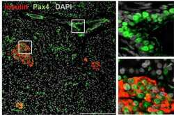
- Immunofluorescence was performed on adult human pancreas tissue. To expose target proteins, antigen retrieval was performed by heating tissues at 95C for 15 minutes in 10mM citrate buffer, pH 6.0. Following antigen retrieval, tissues were blocked and then probed with a Pax4 polyclonal antibody (Product # PA1-108) at a dilution of 1:500 overnight at 4C. Tissues were washed extensively and detection was performed using a fluorescently-conjugated anti-rabbit IgG secondary antibody (green). Tissues were also stained with an antibody against insulin (red), and nuclei (grey) were stained with DAPI. Higher magnification of regions inside the white boxes (left panel) are shown to the right. Scale bar = 200 µm. Data supplied courtesy of the Innovators Program.

- Submitted by
- Invitrogen Antibodies (provider)
- Main image

- Experimental details
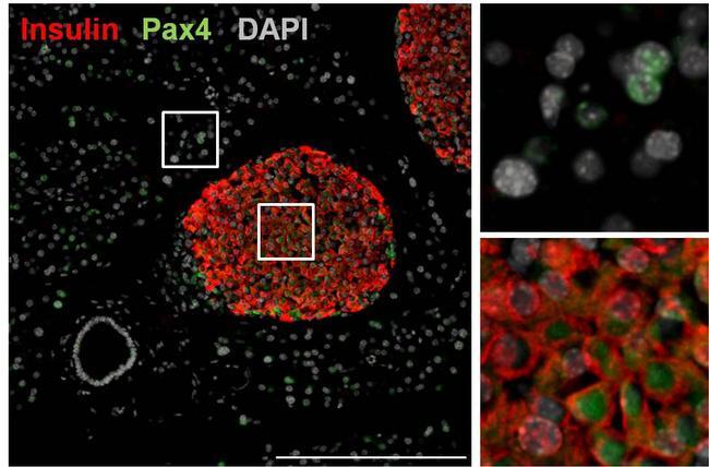
- Immunofluorescence was performed on adult mouse pancreas tissue. To expose target proteins, antigen retrieval was performed by heating tissues at 95C for 15 minutes in 10mM citrate buffer, pH 6.0. Following antigen retrieval, tissues were blocked and then probed with a Pax4 polyclonal antibody (Product # PA1-108) at a dilution of 1:500 overnight at 4C. Tissues were washed extensively and detection was performed using a fluorescently-conjugated anti-rabbit IgG secondary antibody (green). Tissues were also stained with an antibody against insulin (red), and nuclei (grey) were stained with DAPI. Higher magnification of regions inside the white boxes (left panel) are shown to the right. Scale bar = 200 µm. Data supplied courtesy of the Innovators Program.
Supportive validation
- Submitted by
- Invitrogen Antibodies (provider)
- Main image
- Experimental details
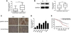
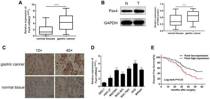
- Figure 1 Elevated PAX4 mRNA and protein expression in both GC tissues and cell lines. ( A , B ) Higher PAX4 mRNA and protein expression in GC tissues compared to normal tissues ( n = 60) by qRT-PCR ( P < 0.0001) and WB ( P = 0.0002). ( C ) The IHC results between GC and non-GC tissues demonstrated excessive PAX4 in GC tissues. ( D ) Overexpression of PAX4 mRNA levels were identified across six GC cell lines (HGC-27, MGC803, BGC-823, SGC-7901, AGS, MKN45) compared to the human gastric mucosal epithelial cell line GES-1. ( E ) The overall survival analysis results demonstrated that PAX4 high expression in GC is associated with poor prognosis.
- Submitted by
- Invitrogen Antibodies (provider)
- Main image

- Experimental details
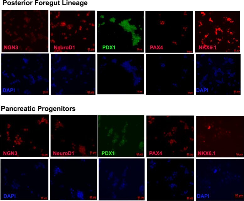
- Fig. 2 Stage wise Immunohistochemistry of the hiPSC cells undergoing differentiation. Foregut lineage cells show positive staining for PDX1 and faint staining for few progenitor markers. Pancreatic progenitor cells show positive staining for all the stage specific markers