Explore
 Validate
 Learn
Learn Western blot
 Immunohistochemistry
Antibody data
- Antibody Data
- Antigen structure
- References [2]
- Comments [0]
- Validations
- Immunohistochemistry [1]
Submit
Validation data
Reference
Comment
Report error
- Product number
- PA5-36153 - Provider product page

- Provider
- Invitrogen Antibodies
- Product name
- UBF-1 Polyclonal Antibody
- Antibody type
- Polyclonal
- Antigen
- Synthetic peptide
- Description
- This antibody detects endogenous protein at a molecular weight of 90 kDa. Purity is >95% by SDS-PAGE.
- Reactivity
- Human, Mouse, Rat
- Host
- Rabbit
- Isotype
- IgG
- Vial size
- 100 μL
- Concentration
- 1 mg/mL
- Storage
- Store at 4°C short term. For long term storage, store at -20°C, avoiding freeze/thaw cycles.
Submitted references Muscle from aged rats is resistant to mechanotherapy during atrophy and reloading.
Massage as a mechanotherapy promotes skeletal muscle protein and ribosomal turnover but does not mitigate muscle atrophy during disuse in adult rats.
Lawrence MM, Van Pelt DW, Confides AL, Hettinger ZR, Hunt ER, Reid JJ, Laurin JL, Peelor FF 3rd, Butterfield TA, Miller BF, Dupont-Versteegden EE
GeroScience 2021 Feb;43(1):65-83
GeroScience 2021 Feb;43(1):65-83
Massage as a mechanotherapy promotes skeletal muscle protein and ribosomal turnover but does not mitigate muscle atrophy during disuse in adult rats.
Lawrence MM, Van Pelt DW, Confides AL, Hunt ER, Hettinger ZR, Laurin JL, Reid JJ, Peelor FF 3rd, Butterfield TA, Dupont-Versteegden EE, Miller BF
Acta physiologica (Oxford, England) 2020 Jul;229(3):e13460
Acta physiologica (Oxford, England) 2020 Jul;229(3):e13460
No comments: Submit comment
Supportive validation
- Submitted by
- Invitrogen Antibodies (provider)
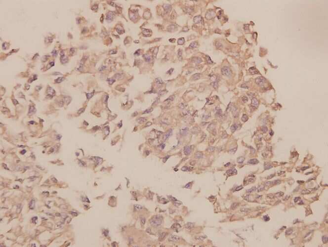
- Main image
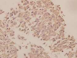
- Experimental details
- Immunohistochemical analysis of UBF-1 in paraffin-embedded human colorectal carcinoma using UBF-1 polyclonal antibody (Product # PA5-36153) at a dilution of 1:50.