 Explore
 Validate
 Learn
Learn Western blot
 Immunohistochemistry
Antibody data
- Antibody Data
- Antigen structure
- References [4]
- Comments [0]
- Validations
- Western blot [1]
- Immunocytochemistry [2]
Submit
Validation data
Reference
Comment
Report error
- Product number
- HPA006385 - Provider product page

- Provider
- Atlas Antibodies
- Proper citation
- Atlas Antibodies Cat#HPA006385, RRID:AB_1080447
- Product name
- Anti-UBTF
- Antibody type
- Polyclonal
- Description
- Polyclonal Antibody against Human UBTF, Gene description: upstream binding transcription factor, RNA polymerase I, Alternative Gene Names: NOR-90, UBF, UBF1, UBF2, Validated applications: ICC, IHC, WB, Uniprot ID: P17480, Storage: Store at +4°C for short term storage. Long time storage is recommended at -20°C.
- Reactivity
- Human, Mouse
- Host
- Rabbit
- Conjugate
- Unconjugated
- Isotype
- IgG
- Vial size
- 100 µl
- Concentration
- 0.1 mg/ml
- Storage
- Store at +4°C for short term storage. Long time storage is recommended at -20°C.
- Handling
- The antibody solution should be gently mixed before use.
Submitted references
Nucleolar Protein Treacle Is Important for the Efficient Growth of Mumps Virus
RNA helicase, DDX27 regulates skeletal muscle growth and regeneration by modulation of translational processes
UBF complexes with phosphatidylinositol 4,5-bisphosphate in nucleolar organizer regions regardless of ongoing RNA polymerase I activity
Arnold M, Cohn G, Oxe K, Elliott S, Moore C, Laraia P, Shekoohi S, Brownell D, Meshul C, Witt S, Larsen D, Unni V
2024
2024
Nucleolar Protein Treacle Is Important for the Efficient Growth of Mumps Virus
Wakata A, Katoh H, Kato F, Takeda M, Dutch R
Journal of Virology 2022;96(19)
Journal of Virology 2022;96(19)
RNA helicase, DDX27 regulates skeletal muscle growth and regeneration by modulation of translational processes
Cox G, Bennett A, O’Donohue M, Gundry S, Chan A, Widrick J, Draper I, Chakraborty A, Zhou Y, Zon L, Gleizes P, Beggs A, Gupta V
PLOS Genetics 2018;14(3):e1007226
PLOS Genetics 2018;14(3):e1007226
UBF complexes with phosphatidylinositol 4,5-bisphosphate in nucleolar organizer regions regardless of ongoing RNA polymerase I activity
Sobol M, Yildirim S, Philimonenko V, Marášek P, Castaño E, Hozák P
Nucleus 2014;4(6):478-486
Nucleus 2014;4(6):478-486
No comments: Submit comment
Enhanced validation
- Submitted by
- Atlas Antibodies (provider)
- Enhanced method
- Genetic validation
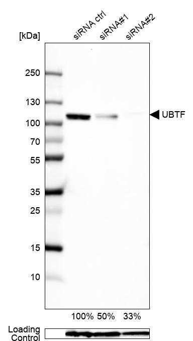
- Main image

- Experimental details
- Western blot analysis in U2OS cells transfected with control siRNA, target specific siRNA probe #1 and #2, using Anti-UBTF antibody. Remaining relative intensity is presented. Loading control: Anti-GAPDH.
- Sample type
- Human
- Protocol
- Protocol
Enhanced validation
Supportive validation
- Submitted by
- 55af80e3e0991
- Enhanced method
- Genetic validation
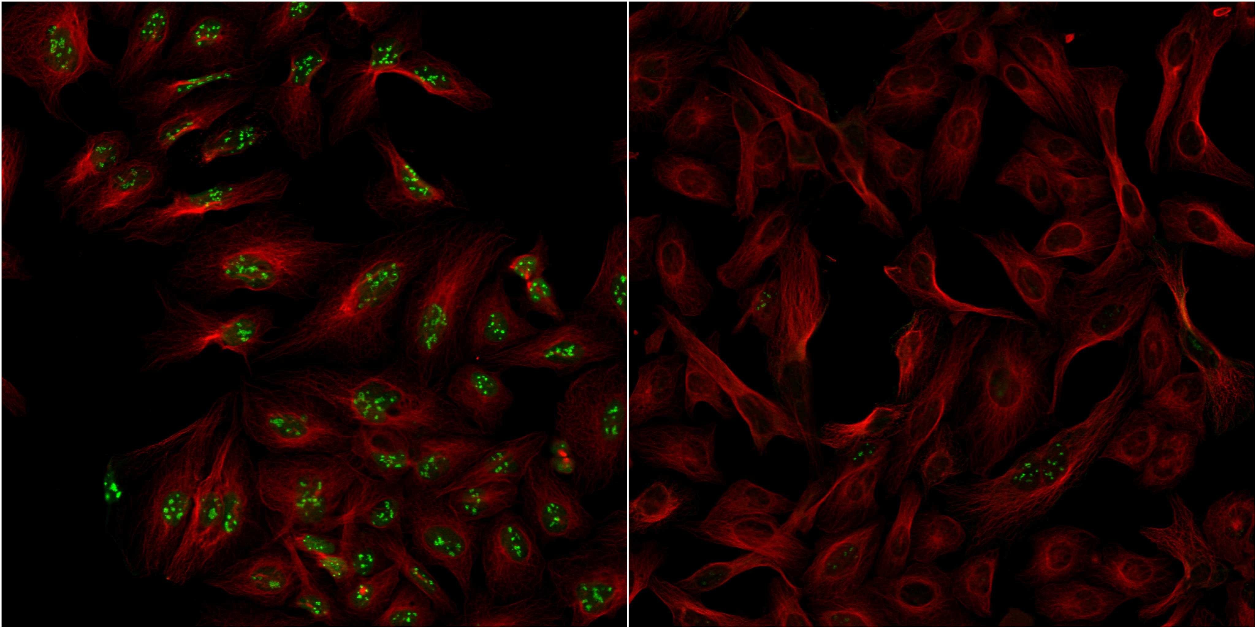
- Main image
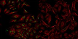
- Experimental details
- Confocal images of immunofluorescently stained human U-2 OS cells.The protein UBTF is shown in green and the microtubules in red. The image to the left show cells transfected with control siRNA and the image to the right show cells where UBTF has been downregulated with specific siRNA.
- Sample type
- U-2 OS cells
- Primary Ab dilution
- 1:72
- Secondary Ab
- Secondary Ab
- Secondary Ab dilution
- 1:800
- Knockdown/Genetic Approaches Application
- Immunocytochemistry
Supportive validation
- Submitted by
- Atlas Antibodies (provider)
- Main image
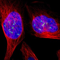
- Experimental details
- Immunofluorescent staining of human cell line U-2 OS shows localization to nucleoli fibrillar center.
- Sample type
- Human
