Explore
 Validate
 Learn
Learn Western blot
Antibody data
- Antibody Data
- Antigen structure
- References [1]
- Comments [0]
- Validations
- Western blot [3]
- Immunocytochemistry [2]
- Immunohistochemistry [1]
Submit
Validation data
Reference
Comment
Report error
- Product number
- GTX102061 - Provider product page

- Provider
- GeneTex
- Proper citation
- GeneTex Cat#GTX102061, RRID:AB_2037187
- Product name
- hnRNP H antibody [N1C1]
- Antibody type
- Polyclonal
- Reactivity
- Human, Mouse
- Host
- Rabbit
Submitted references 3'poly-G-tailed ODNs inhibit F-spondin to induce cell death and neurite retraction in rat embryonic neurons.
Cheng YC, Chen TA, Chen CY, Liang CM, Liang SM
Molecular neurobiology 2012 Jun;45(3):536-49
Molecular neurobiology 2012 Jun;45(3):536-49
No comments: Submit comment
Supportive validation
- Submitted by
- GeneTex (provider)
- Main image
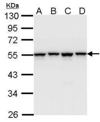
- Experimental details
- Sample (30 ug of whole cell lysate) A: A431 (GTX27909) B: H1299 C: Hela D: Hep G2 (GTX27900) 10% SDS PAGE GTX102061 diluted at 1:1000

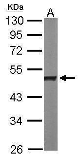
- Submitted by
- GeneTex (provider)
- Main image

- Experimental details
- Sample (50 ug of whole cell lysate) A: Mouse brain 10% SDS PAGE GTX102061 diluted at 1:1000
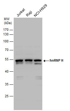
- Submitted by
- GeneTex (provider)
- Main image
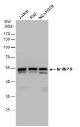
- Experimental details
- hnRNP H antibody detects hnRNP H protein by western blot analysis. Various whole cell extracts (30 £gg) were separated by 10% SDS-PAGE, and the membrane was blotted with hnRNP H antibody (GTX102061) diluted by 1:1000.
Supportive validation
- Submitted by
- GeneTex (provider)
- Main image
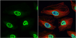
- Experimental details
- hnRNP H antibody [N1C1] detects hnRNP H protein at nucleus by immunofluorescent analysis.Sample: HeLa cells were fixed in 4% paraformaldehyde at RT for 15 min.Green: hnRNP H protein stained by hnRNP H antibody [N1C1] (GTX102061) diluted at 1:200.Red: alpha Tubulin, a cytoskeleton marker, stained by alpha Tubulin antibody [B-5-1-2] (GTX11304) diluted at 1:10000.Blue: Hoechst 33342 staining.

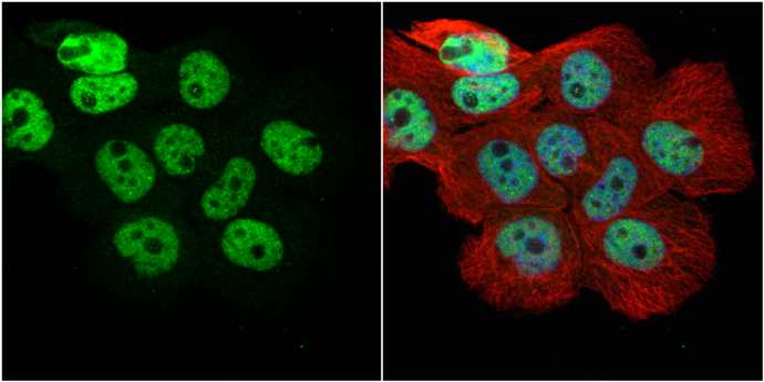
- Submitted by
- GeneTex (provider)
- Main image

- Experimental details
- hnRNP H antibody [N1C1] detects hnRNP H protein at nucleus by immunofluorescent analysis.Sample: A431 cells were fixed in 4% paraformaldehyde at RT for 15 min.Green: hnRNP H protein stained by hnRNP H antibody [N1C1] (GTX102061) diluted at 1:500.Red: alpha Tubulin, a cytoskeleton marker, stained by alpha Tubulin antibody [GT114] (GTX628802) diluted at 1:1000.Blue: Hoechst 33342 staining.
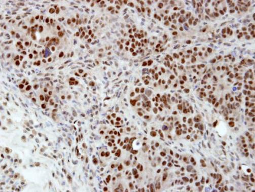
Supportive validation
- Submitted by
- GeneTex (provider)
- Main image
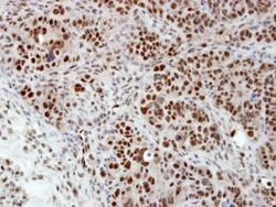
- Experimental details
- Immunohistochemical analysis of paraffin-embedded NCIN87 xenograft, using hnRNP H(GTX102061) antibody at 1:100 dilution.