Explore
 Validate
 Learn
Learn Western blot
Antibody data
- Antibody Data
- Antigen structure
- References [1]
- Comments [0]
- Validations
- Western blot [3]
- Immunocytochemistry [5]
- Immunohistochemistry [2]
- Other assay [1]
Submit
Validation data
Reference
Comment
Report error
- Product number
- PA5-27610 - Provider product page

- Provider
- Invitrogen Antibodies
- Product name
- hnRNP H1 Polyclonal Antibody
- Antibody type
- Polyclonal
- Antigen
- Recombinant full-length protein
- Description
- Recommended positive controls: A431, H1299, HeLa, HepG2, Raji, NCI-H929, mouse brain. Predicted reactivity: Mouse (100%), Rat (99%), Zebrafish (87%), Xenopus laevis (91%), Chicken (96%), Bovine (100%). Store product as a concentrated solution. Centrifuge briefly prior to opening the vial.
- Reactivity
- Human, Mouse
- Host
- Rabbit
- Isotype
- IgG
- Vial size
- 100 μL
- Concentration
- 1 mg/mL
- Storage
- Store at 4°C short term. For long term storage, store at -20°C, avoiding freeze/thaw cycles.
Submitted references Unexpected similarities between C9ORF72 and sporadic forms of ALS/FTD suggest a common disease mechanism.
Conlon EG, Fagegaltier D, Agius P, Davis-Porada J, Gregory J, Hubbard I, Kang K, Kim D, New York Genome Center ALS Consortium, Phatnani H, Shneider NA, Manley JL
eLife 2018 Jul 13;7
eLife 2018 Jul 13;7
No comments: Submit comment
Supportive validation
- Submitted by
- Invitrogen Antibodies (provider)
- Main image
- Experimental details
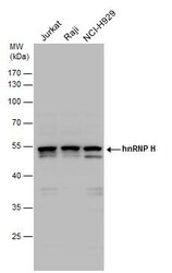
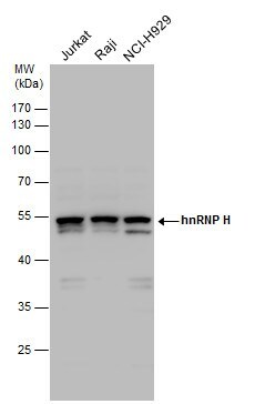
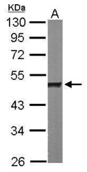
- hnRNP H antibody detects hnRNP H protein by western blot analysis. Various whole cell extracts (30 µg) were separated by 10% SDS-PAGE, and the membrane was blotted with hnRNP H antibody hnRNP H1 Polyclonal Antibody (Product # PA5-27610) diluted by 1:1,000.
- Submitted by
- Invitrogen Antibodies (provider)
- Main image
- Experimental details
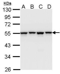
- Western Blot using hnRNP H1 Polyclonal Antibody (Product # PA5-27610). Sample (30 µg of whole cell lysate). Lane A: A431. Lane B: H1299. Lane C: Hela. Lane D: Hep G2. 10% SDS PAGE. hnRNP H1 Polyclonal Antibody (Product # PA5-27610) diluted at 1:1,000.

- Submitted by
- Invitrogen Antibodies (provider)
- Main image
- Experimental details
- Western Blot using hnRNP H1 Polyclonal Antibody (Product # PA5-27610). Sample (50 µg of whole cell lysate). Lane A: Mouse brain. 10% SDS PAGE. hnRNP H1 Polyclonal Antibody (Product # PA5-27610) diluted at 1:1,000.

Supportive validation
- Submitted by
- Invitrogen Antibodies (provider)
- Main image
- Experimental details
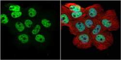
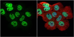
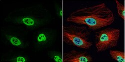
- Immunocytochemistry-Immunofluorescence analysis of hnRNP H1 was performed in A431 cells fixed in 4% paraformaldehyde at RT for 15 min. Green: hnRNP H1 Polyclonal Antibody (Product # PA5 27610) diluted at 1:500. Red: alpha Tubulin, a cytoskeleton marker. Blue: Hoechst 33342 staining.
- Submitted by
- Invitrogen Antibodies (provider)
- Main image

- Experimental details
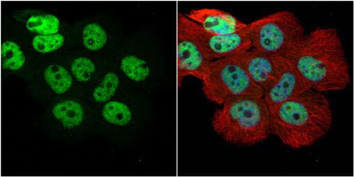
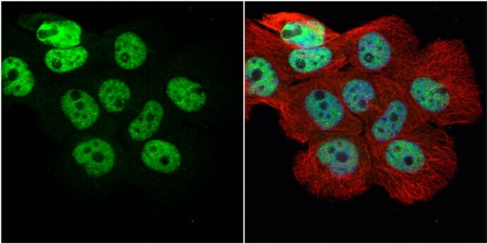
- Immunocytochemistry-Immunofluorescence analysis of hnRNP H1 was performed in HeLa cells fixed in 4% paraformaldehyde at RT for 15 min. Green: hnRNP H1 Polyclonal Antibody (Product # PA5 27610) diluted at 1:200. Red: alpha Tubulin, a cytoskeleton marker. Blue: Hoechst 33342 staining.

- Submitted by
- Invitrogen Antibodies (provider)
- Main image
- Experimental details
- Immunocytochemistry-Immunofluorescence analysis of hnRNP H1 was performed in A431 cells fixed in 4% paraformaldehyde at RT for 15 min. Green: hnRNP H1 Polyclonal Antibody (Product # PA5 27610) diluted at 1:500. Red: alpha Tubulin, a cytoskeleton marker. Blue: Hoechst 33342 staining.

- Submitted by
- Invitrogen Antibodies (provider)
- Main image
- Experimental details
- Immunocytochemistry-Immunofluorescence analysis of hnRNP H1 was performed in HeLa cells fixed in 4% paraformaldehyde at RT for 15 min. Green: hnRNP H1 Polyclonal Antibody (Product # PA5 27610) diluted at 1:200. Red: alpha Tubulin, a cytoskeleton marker. Blue: Hoechst 33342 staining.

- Submitted by
- Invitrogen Antibodies (provider)
- Main image

- Experimental details
- Immunocytochemistry-Immunofluorescence analysis of hnRNP H1 was performed in A431 cells fixed in 4% paraformaldehyde at RT for 15 min. Green: hnRNP H1 Polyclonal Antibody (Product # PA5 27610) diluted at 1:500. Red: alpha Tubulin, a cytoskeleton marker. Blue: Hoechst 33342 staining.
Supportive validation
- Submitted by
- Invitrogen Antibodies (provider)
- Main image
- Experimental details
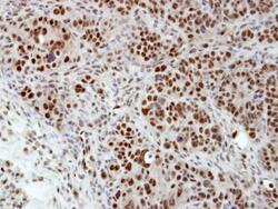
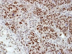
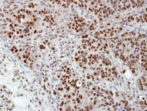
- Immunohistochemical analysis of paraffin-embedded NCIN87 xenograft, using hnRNP H (Product # PA5-27610) antibody at 1:100 dilution. Antigen Retrieval: EDTA based buffer, pH 8.0, 15 min.
- Submitted by
- Invitrogen Antibodies (provider)
- Main image
- Experimental details
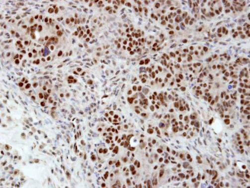
- Immunohistochemical analysis of paraffin-embedded NCIN87 xenograft, using hnRNP H (Product # PA5-27610) antibody at 1:100 dilution. Antigen Retrieval: EDTA based buffer, pH 8.0, 15 min.
Supportive validation
- Submitted by
- Invitrogen Antibodies (provider)
- Main image

- Experimental details
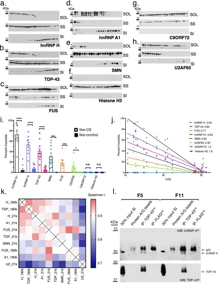
- Figure 4. Like-control ALS/FTD patients have undetectable levels of insoluble RBPs after low-speed fractionation. ( a-h ) Low-speed (21,000 x G) fractionation experiment with 20 patients listed in Figure 3a . Western blot hnRNP H ( a ), TDP-43 ( b ), FUS ( c ), hnRNP A1 ( d ), SMN ( e ), Histone H3 ( f ), C9ORF72 ( g ), U2AF65 ( h ). SOL = soluble, SS = sarkosyl soluble, SI = sarkosyl insoluble. ( i ) Quantification of low-speed centrifugation of 20 patients with replicate values. Error bars are plotted to the SEM. ( j ) Linear regression of percent insoluble protein for targets in ( a-h ) plotted against CSS. R-square is listed. ( k ) Heatmap of spearman correlation coefficients for each pairwise comparison of percent insoluble protein at 180,000 or 21,000 x G (180 k and21k, respectively). ( l ) Western blots of hnRNP H (top) and TDP-43 (bottom) following IP with protein A/G magnetic beads alone, TDP-43 antibody, or control (FLAG) antibody from two patient brains (F5 and F11). The abbreviations ms and rb stand for mouse antibody and rabbit antibody, respectively. Protein species are indicated by arrows at right, and positions of size markers are shown on the left. Figure 4--figure supplement 1. Insoluble hnRNP H and TDP-43 correlate in C9 patient brains. Representative western blot of 21,000 x G motor cortex fractionation from eight controls (six nonALS and two SOD1 ALS) and 14 C9+ ALS/FTD patients. Membranes are shown from top-bottom: cortex homogenate (western blot actin),