Explore
 Validate
 Learn
Learn Western blot
 Immunohistochemistry
Antibody data
- Antibody Data
- Antigen structure
- References [1]
- Comments [0]
- Validations
- Immunohistochemistry [1]
- Other assay [1]
Submit
Validation data
Reference
Comment
Report error
- Product number
- PA5-93062 - Provider product page

- Provider
- Invitrogen Antibodies
- Product name
- COL9A1 Polyclonal Antibody
- Antibody type
- Polyclonal
- Antigen
- Recombinant full-length protein
- Reactivity
- Human, Rat
- Host
- Rabbit
- Isotype
- IgG
- Vial size
- 100 µL
- Concentration
- 0.32 mg/mL
- Storage
- -20° C, Avoid Freeze/Thaw Cycles
Submitted references Noise Exposures Causing Hearing Loss Generate Proteotoxic Stress and Activate the Proteostasis Network.
Jongkamonwiwat N, Ramirez MA, Edassery S, Wong ACY, Yu J, Abbott T, Pak K, Ryan AF, Savas JN
Cell reports 2020 Nov 24;33(8):108431
Cell reports 2020 Nov 24;33(8):108431
No comments: Submit comment
Supportive validation
- Submitted by
- Invitrogen Antibodies (provider)
- Main image
- Experimental details
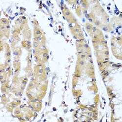
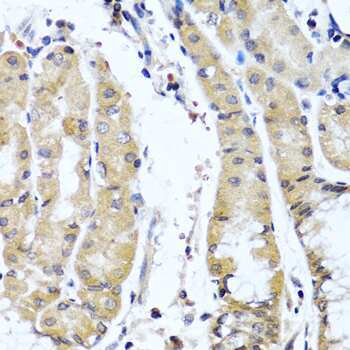
- Immunohistochemistry analysis of COL9A1 in paraffin-embedded human stomach using COL9A1 Polyclonal Antibody (Product # PA5-93062) at a dilution of 1:200.
Supportive validation
- Submitted by
- Invitrogen Antibodies (provider)
- Main image
- Experimental details
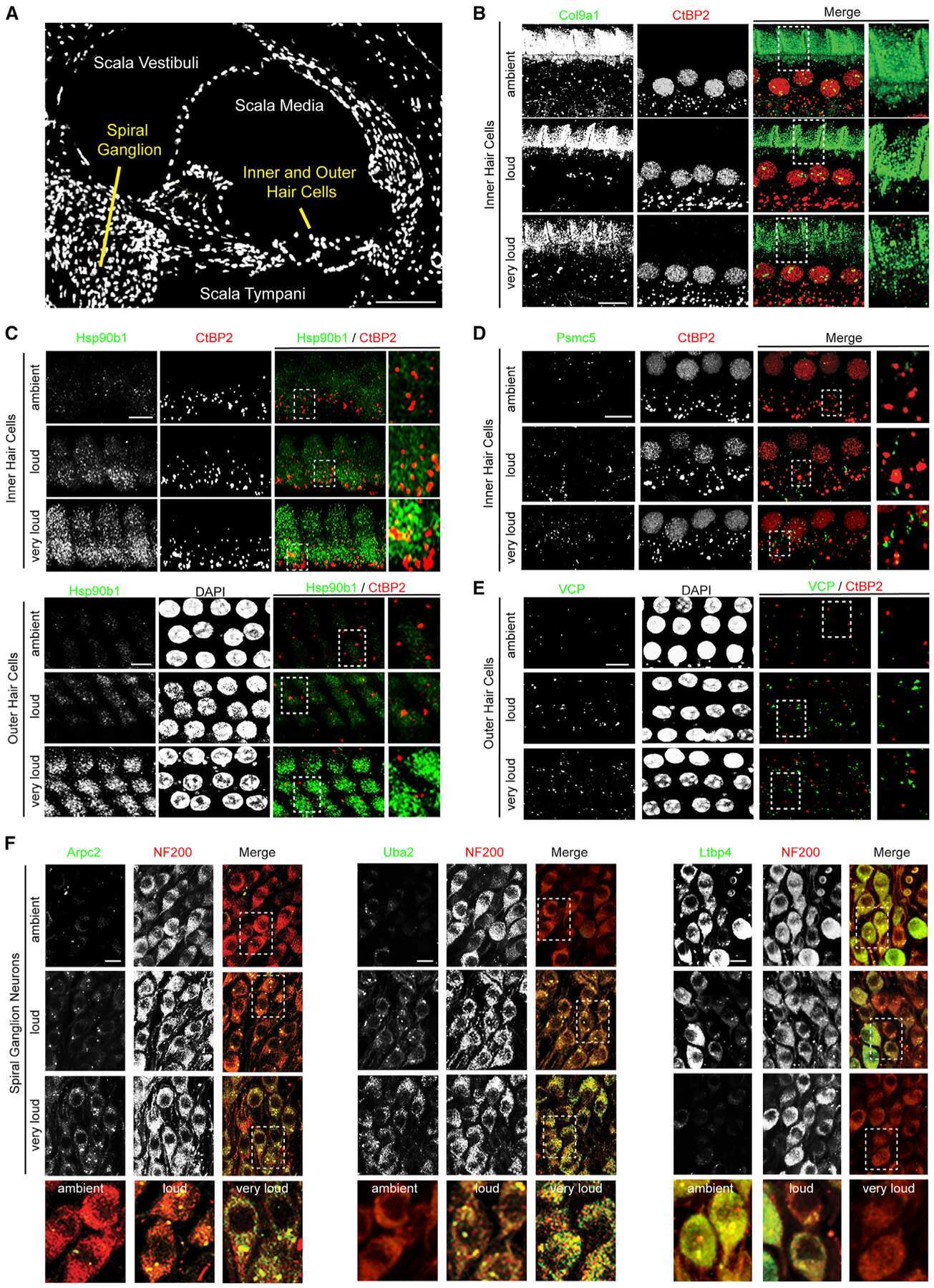
- Figure 4. Proteins Regulated during Noise Exposure Are Expressed by HCs, SGNs, and Supporting Cells within the Cochlea (A) DAPI-stained midmodiolar cochlear section. (B) Col9a1 signal decreases in the area above the tunnel of Corti following noise exposure. Green, Col9a1; red, CtBP2. (C) Hsp90b1 levels are elevated in both IHCs and OHCs after exposure to increasing intensities of noise. Green, Hsp90b1; red, CtBP2; white, DAPI. (D) Psmc5 levels increase after exposure to increasing noise intensity within IHCs. Green, Psmc5; red, CtBP2. (E) Vcp levels are elevated in the OHCs after exposure to increasingly damaging levels of noise. Green, Vcp; red, CtBP2; white, DAPI. (F) SGNs from the second ganglion bundle from the top of the cochlea (12-16 kHz) in midmodiolar sections. Left: Arpc2 levels are elevated in SGNs after exposure to increasingly damaging levels of noise. Middle: Uba2 levels progressively increase within SGNs with increasing noise exposure. Right: LtBP4 levels are reduced in SGNs after exposure to increasingly damaging levels of noise. Green, Arpc2, Uba2, and LtBP4 (left, middle, and right); red, NF200. Scale bars, 100 mum (A) and 10 mum (B-F). Representative images from n = 2 animals.