Explore
 Validate
 Learn
Learn Western blot
Antibody data
- Antibody Data
- Antigen structure
- References [0]
- Comments [0]
- Validations
- Western blot [4]
- Immunohistochemistry [3]
Submit
Validation data
Reference
Comment
Report error
- Product number
- PA5-20004 - Provider product page

- Provider
- Invitrogen Antibodies
- Product name
- ENDOG Polyclonal Antibody
- Antibody type
- Polyclonal
- Antigen
- Synthetic peptide
- Description
- In Western blot applications, this antibody detects a band at ~35kDa. A suggested positive control is HepG2 cell lysate.
- Concentration
- 1 mg/mL
No comments: Submit comment
Supportive validation
- Submitted by
- Invitrogen Antibodies (provider)
- Main image
- Experimental details
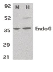
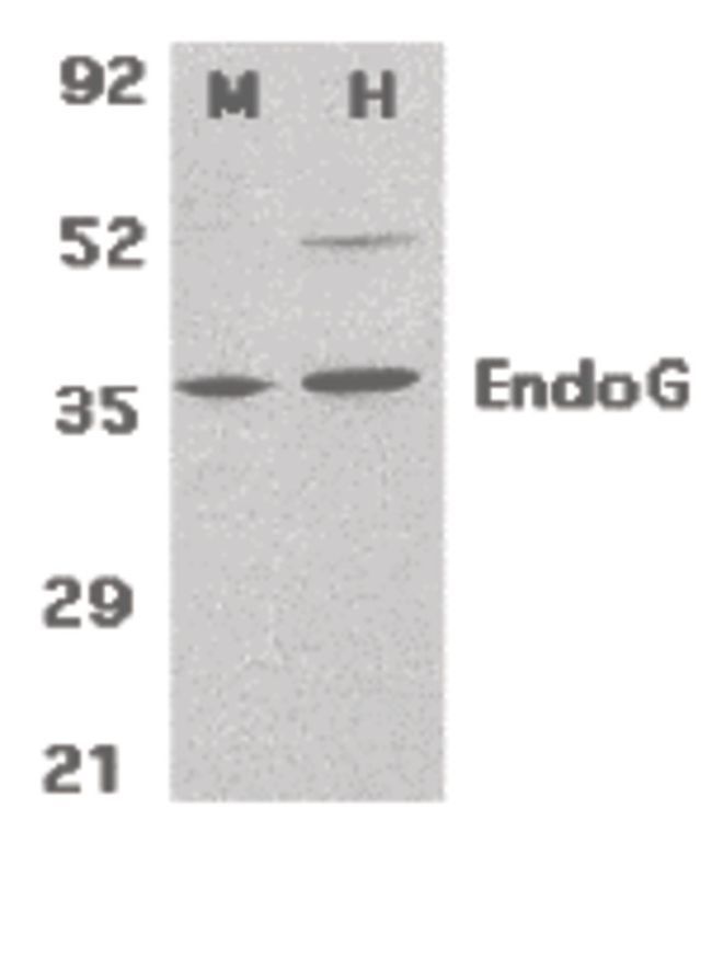
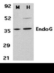
- Western blot analysis of mouse (M) 3T3 and human (H) HepG2 cell lysates using a EndoG polyclonal antibody (Product # PA5-20004) at 2 µg/mL.
- Submitted by
- Invitrogen Antibodies (provider)
- Main image

- Experimental details
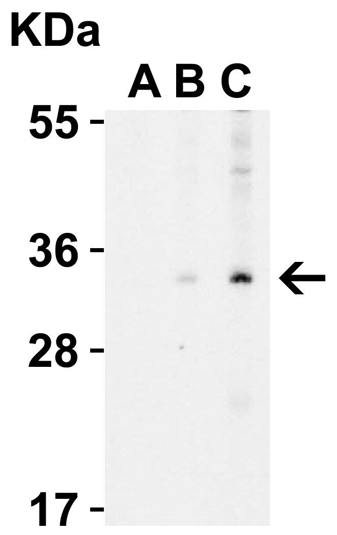
- Western Blot Validation in Human A431 Cell Lysate with the presence (A) or absence (B and C) of blocking peptide. Loading: 15 µg of lysates per lane. Antibodies: ENDOG Polyclonal Antibody (Product # PA5-20004) (A: 0.5 µg/mL, B: 0.5 µg/mL, C: 1 µg/mL), 1h incubation at RT in 0.05 NFDM/TBST. Secondary: Goat anti-rabbit IgG HRP conjugate at 1:10,000 dilution.
- Submitted by
- Invitrogen Antibodies (provider)
- Main image
- Experimental details
- Western Blot Validation in Mouse 3T3 (M) and Human HepG2 (H) Cell Lysates. Loading: 15 µg of lysates per lane. Antibodies: ENDOG Polyclonal Antibody (Product # PA5-20004) (2 µg/mL), 1h incubation at RT in 0.05 NFDM/TBST. Secondary: Goat anti-rabbit IgG HRP conjugate at 1:10,000 dilution.

- Submitted by
- Invitrogen Antibodies (provider)
- Main image
- Experimental details
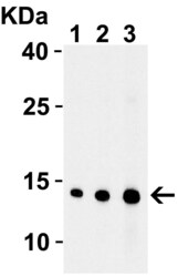
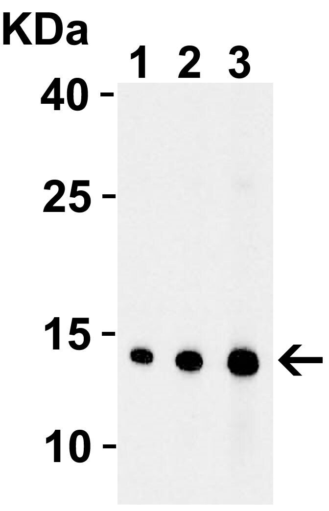
- Western Blot Validation with Recombinant Protein. Loading: 30 ng of human EndoG recombinant protein per lane. Antibodies: ENDOG Polyclonal Antibody (Product # PA5-20004) 1h incubation at RT in 0.05 NFDM/TBST. Secondary: Goat anti-rabbit IgG HRP conjugate at 1:10,000 dilution. Lane 1: 0.5 µg/mL Lane 2: 1 µg/mL Lane 3: 2 µg/mL.
Supportive validation
- Submitted by
- Invitrogen Antibodies (provider)
- Main image

- Experimental details
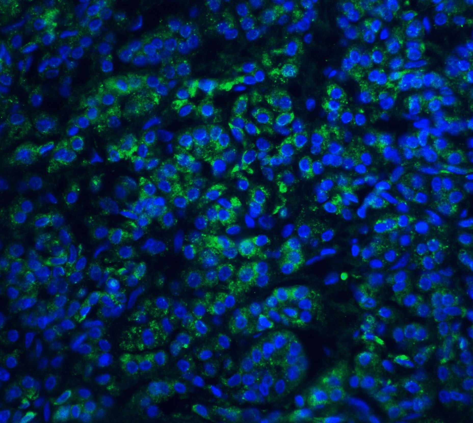
- Immunofluorescent analysis of 4% paraformaldehyde-fixed human pancreas tissue labeling EndoG with ENDOG Polyclonal Antibody (Product # PA5-20004) at 20 µg/mL, followed by goat anti-rabbit IgG secondary antibody at 1:500 dilution (green) and DAPI staining (blue).
- Submitted by
- Invitrogen Antibodies (provider)
- Main image
- Experimental details
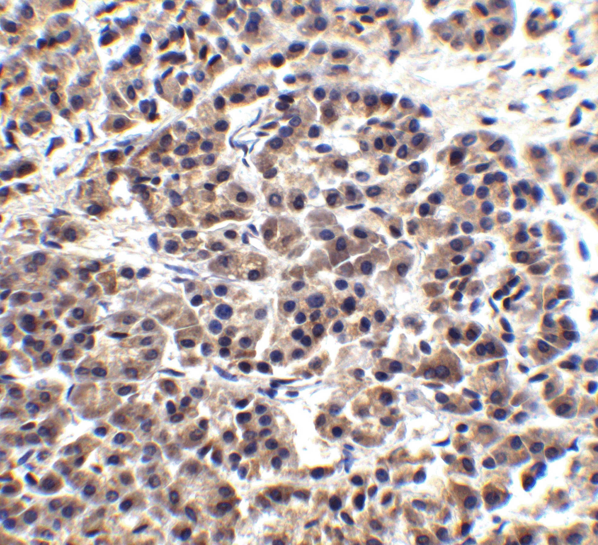
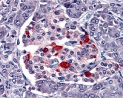
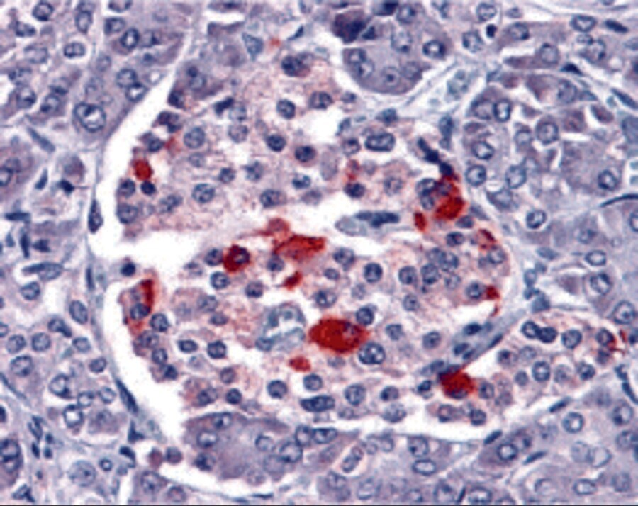
- Immunohistochemical analysis of paraffin-embedded human pancreas tissue using ENDOG Polyclonal Antibody (Product # PA5-20004) at 15 µg/mL. Tissue was fixed with formaldehyde and blocked with 0.1 serum for 1 h at RT; antigen retrieval was by heat mediation with a citrate buffer (pH6). Samples were incubated with primary antibody overnight at 4˚C. A goat anti-rabbit IgG H&L (HRP) at 1/250 was used as secondary. Counter stained with Hematoxylin.
- Submitted by
- Invitrogen Antibodies (provider)
- Main image

- Experimental details
- Immunohistochemical analysis of paraffin-embedded human pancreas tissue using ENDOG Polyclonal Antibody (Product # PA5-20004) at 2.5 µg/mL. Tissue was fixed with formaldehyde and blocked with 0.1 serum for 1 h at RT; antigen retrieval was by heat mediation with a citrate buffer (pH6). Samples were incubated with primary antibody overnight at 4˚C. A goat anti-rabbit IgG H&L (HRP) at 1/250 was used as secondary. Counter stained with Hematoxylin.