 Explore
 Validate
 Learn
Learn Western blot
 Immunocytochemistry
 Immunohistochemistry
Antibody data
- Antibody Data
- Antigen structure
- References [4]
- Comments [0]
- Validations
- Western blot [1]
- Immunocytochemistry [1]
Submit
Validation data
Reference
Comment
Report error
- Product number
- HPA021323 - Provider product page

- Provider
- Atlas Antibodies
- Proper citation
- Atlas Antibodies Cat#HPA021323, RRID:AB_1849554
- Product name
- Anti-GCC1
- Antibody type
- Polyclonal
- Description
- Polyclonal Antibody against Human GCC1, Gene description: GRIP and coiled-coil domain containing 1, Alternative Gene Names: FLJ22035, GCC1P, GCC88, MGC20706, Validated applications: ICC, IHC, WB, Uniprot ID: Q96CN9, Storage: Store at +4°C for short term storage. Long time storage is recommended at -20°C.
- Reactivity
- Human, Mouse, Rat
- Host
- Rabbit
- Conjugate
- Unconjugated
- Isotype
- IgG
- Vial size
- 100 µl
- Concentration
- 0.2 mg/ml
- Storage
- Store at +4°C for short term storage. Long time storage is recommended at -20°C.
- Handling
- The antibody solution should be gently mixed before use.
Submitted references ARFRP1 functions upstream of ARL1 and ARL5 to coordinate recruitment of distinct tethering factors to the trans-Golgi network.
The golgin coiled-coil proteins capture different types of transport carriers via distinct N-terminal motifs
MiR-199a Inhibits Secondary Envelopment of Herpes Simplex Virus-1 Through the Downregulation of Cdc42-specific GTPase Activating Protein Localized in Golgi Apparatus.
The specificity of vesicle traffic to the Golgi is encoded in the golgin coiled-coil proteins
Ishida M, Bonifacino JS
The Journal of cell biology 2019 Nov 4;218(11):3681-3696
The Journal of cell biology 2019 Nov 4;218(11):3681-3696
The golgin coiled-coil proteins capture different types of transport carriers via distinct N-terminal motifs
Wong M, Gillingham A, Munro S
BMC Biology 2017;15(1)
BMC Biology 2017;15(1)
MiR-199a Inhibits Secondary Envelopment of Herpes Simplex Virus-1 Through the Downregulation of Cdc42-specific GTPase Activating Protein Localized in Golgi Apparatus.
Kobayashi K, Suemasa F, Sagara H, Nakamura S, Ino Y, Kobayashi K, Hiramatsu H, Haraguchi T, Kurokawa K, Todo T, Nakano A, Iba H
Scientific reports 2017 Jul 27;7(1):6650
Scientific reports 2017 Jul 27;7(1):6650
The specificity of vesicle traffic to the Golgi is encoded in the golgin coiled-coil proteins
Wong M, Munro S
Science 2014;346(6209)
Science 2014;346(6209)
No comments: Submit comment
Enhanced validation
- Submitted by
- Atlas Antibodies (provider)
- Enhanced method
- Genetic validation
- Main image
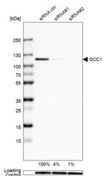
- Experimental details
- Western blot analysis in U-138MG cells transfected with control siRNA, target specific siRNA probe #1 and #2, using Anti-GCC1 antibody. Remaining relative intensity is presented. Loading control: Anti-GAPDH.
- Sample type
- Human
- Protocol
- Protocol

Supportive validation
- Submitted by
- Atlas Antibodies (provider)
- Main image
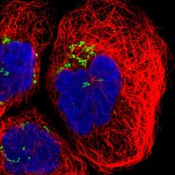
- Experimental details
- Immunofluorescent staining of human cell line A-431 shows localization to the Golgi apparatus.
- Sample type
- Human
