 Explore
 Validate
 Learn
Learn Western blot
Antibody data
- Antibody Data
- Antigen structure
- References [1]
- Comments [0]
- Validations
- Western blot [2]
- Immunocytochemistry [1]
- Immunohistochemistry [4]
Submit
Validation data
Reference
Comment
Report error
- Product number
- PA5-53304 - Provider product page

- Provider
- Invitrogen Antibodies
- Product name
- NOX4 Polyclonal Antibody
- Antibody type
- Polyclonal
- Antigen
- Recombinant full-length protein
- Description
- Immunogen sequence: ISLNRTSSQN ISLPEYFSEH FHEPFPEGFS KPAEFTQHKF VKICMEEPRF QANFPQTWLW ISGPLCLYCA ERLYRYIRSN KPVTIISVIS HPSDVMEIRM VKENFKARPG QYI
- Concentration
- 0.1 mg/mL
Submitted references Setanaxib (GKT137831) Ameliorates Doxorubicin-Induced Cardiotoxicity by Inhibiting the NOX1/NOX4/Reactive Oxygen Species/MAPK Pathway.
Zheng H, Xu N, Zhang Z, Wang F, Xiao J, Ji X
Frontiers in pharmacology 2022;13:823975
Frontiers in pharmacology 2022;13:823975
No comments: Submit comment
Supportive validation
- Submitted by
- Invitrogen Antibodies (provider)
- Main image
- Experimental details
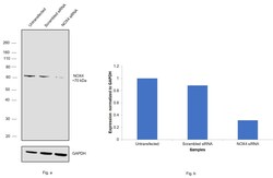
- Knockdown of NOX4 was achieved by transfecting A549 with NOX4 specific siRNAs (Silencer® select Product # S224160, S224159). Western blot analysis (Fig. a) was performed using Whole cell extracts from the NOX4 knockdown cells (lane 3), non-targeting scrambled siRNA transfected cells (lane 2) and untransfected cells (lane 1). The blot was probed with NOX4 Polyclonal Antibody (Product # PA5-53304, 1:1000 dilution) and Goat anti-Rabbit IgG (H+L) Superclonal™ Recombinant Secondary Antibody, HRP (Product # A27036, 1:4000 dilution). Densitometric analysis of this western blot is shown in histogram (Fig. b). Decrease in signal upon siRNA mediated knock down confirms that antibody is specific to NOX4.

- Submitted by
- Invitrogen Antibodies (provider)
- Main image

- Experimental details
- Western blot was performed using Anti-NOX4 Polyclonal Antibody (Product # PA5-53304) and a 70kDa band corresponding to NOX4 was observed across in A549 and Mouse Kidney in comparison to NIH: OVCAR-3 which is reported to be negative. Whole cell extracts (30 µg lysate) of A549 (Lane 1), NIH:OVCAR-3 (Lane 2), Mouse Kidney (Lane 3) were electrophoresed using NuPAGE™ 4-12% Bis-Tris Protein Gel (Product # NP0322BOX). Resolved proteins were then transferred onto a Nitrocellulose membrane (Product # IB23001) by iBlot® 2 Dry Blotting System (Product # IB21001). The blot was probed with the primary antibody (1:500 dilution) and detected by chemiluminescence with Goat anti-Rabbit IgG (H+L) Superclonal™ Recombinant Secondary Antibody, HRP (Product # A27036, 1:4000 dilution) using the iBright FL 1000 (Product # A32752). Chemiluminescent detection was performed using Novex® ECL Chemiluminescent Substrate Reagent Kit (Product # WP20005).

Supportive validation
- Submitted by
- Invitrogen Antibodies (provider)
- Main image

- Experimental details
- Immunofluorescent staining of NOX4 in human cell line HUVEC TERT2 using a NOX4 Polyclonal Antibody (Product # PA5-53304) shows localization to nucleus.

Supportive validation
- Submitted by
- Invitrogen Antibodies (provider)
- Main image

- Experimental details
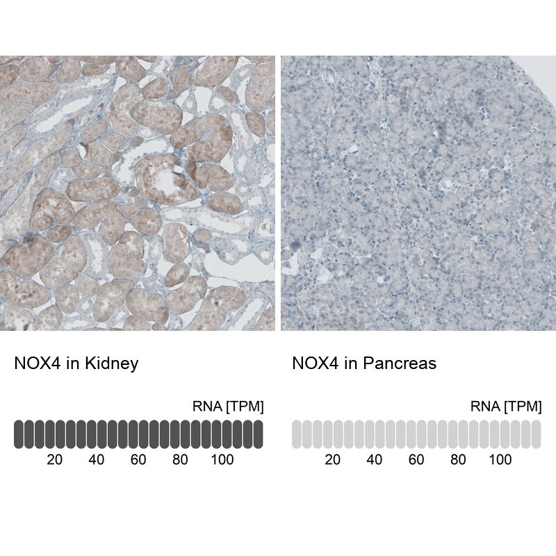
- Immunohistochemical staining of NOX4 in human kidney and pancreas tissues using NOX4 Polyclonal Antibody (Product # PA5-53304). Corresponding NOX4 RNA-seq data are presented for the same tissues.
- Submitted by
- Invitrogen Antibodies (provider)
- Main image

- Experimental details
- Immunohistochemical staining of NOX4 in human kidney using NOX4 Polyclonal Antibody (Product # PA5-53304) shows weak to moderate cytoplasmic positivity in cells in proximal tubules.

- Submitted by
- Invitrogen Antibodies (provider)
- Main image

- Experimental details
- Immunohistochemical staining of NOX4 in human testis using NOX4 Polyclonal Antibody (Product # PA5-53304) shows weak cytoplasmic positivity in cells in seminiferous ducts.

- Submitted by
- Invitrogen Antibodies (provider)
- Main image

- Experimental details
- Immunohistochemical staining of NOX4 in human pancreas using NOX4 Polyclonal Antibody (Product # PA5-53304) shows moderate positivity in exocrine glandular cells.
