Explore
 Validate
 Learn
Learn Western blot
 Immunocytochemistry
 Flow cytometry
Antibody data
- Antibody Data
- Antigen structure
- References [3]
- Comments [0]
- Validations
- Immunocytochemistry [5]
- Immunohistochemistry [4]
- Other assay [3]
Submit
Validation data
Reference
Comment
Report error
- Product number
- MA5-32090 - Provider product page

- Provider
- Invitrogen Antibodies
- Product name
- NOX4 Recombinant Rabbit Monoclonal Antibody (SY0214)
- Antibody type
- Monoclonal
- Antigen
- Recombinant full-length protein
- Description
- Recombinant rabbit monoclonal antibodies are produced using in vitro expression systems. The expression systems are developed by cloning in the specific antibody DNA sequences from immunoreactive rabbits. Then, individual clones are screened to select the best candidates for production. The advantages of using recombinant rabbit monoclonal antibodies include: better specificity and sensitivity, lot-to-lot consistency, animal origin-free formulations, and broader immunoreactivity to diverse targets due to larger rabbit immune repertoire.
- Reactivity
- Human, Mouse, Rat
- Host
- Rabbit
- Isotype
- IgG
- Antibody clone number
- SY0214
- Vial size
- 100 μL
- Concentration
- 1 mg/mL
- Storage
- Store at 4°C short term. For long term storage, store at -20°C, avoiding freeze/thaw cycles.
Submitted references IL11 stimulates ERK/P90RSK to inhibit LKB1/AMPK and activate mTOR initiating a mesenchymal program in stromal, epithelial, and cancer cells.
Hepatocyte-specific IL11 cis-signaling drives lipotoxicity and underlies the transition from NAFLD to NASH.
NOX1 Inhibition Attenuates Kidney Ischemia-Reperfusion Injury via Inhibition of ROS-Mediated ERK Signaling.
Widjaja AA, Viswanathan S, Wei Ting JG, Tan J, Shekeran SG, Carling D, Lim WW, Cook SA
iScience 2022 Aug 19;25(8):104806
iScience 2022 Aug 19;25(8):104806
Hepatocyte-specific IL11 cis-signaling drives lipotoxicity and underlies the transition from NAFLD to NASH.
Dong J, Viswanathan S, Adami E, Singh BK, Chothani SP, Ng B, Lim WW, Zhou J, Tripathi M, Ko NSJ, Shekeran SG, Tan J, Lim SY, Wang M, Lio PM, Yen PM, Schafer S, Cook SA, Widjaja AA
Nature communications 2021 Jan 4;12(1):66
Nature communications 2021 Jan 4;12(1):66
NOX1 Inhibition Attenuates Kidney Ischemia-Reperfusion Injury via Inhibition of ROS-Mediated ERK Signaling.
Jung HY, Oh SH, Ahn JS, Oh EJ, Kim YJ, Kim CD, Park SH, Kim YL, Cho JH
International journal of molecular sciences 2020 Sep 21;21(18)
International journal of molecular sciences 2020 Sep 21;21(18)
No comments: Submit comment
Supportive validation
- Submitted by
- Invitrogen Antibodies (provider)
- Main image

- Experimental details
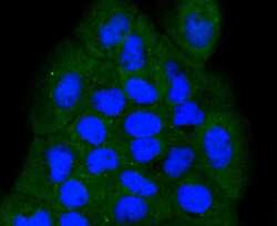
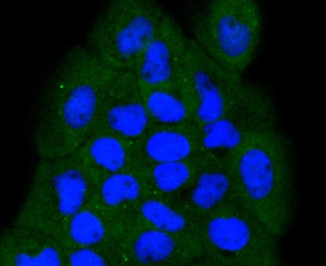
- Immunocytochemical analysis of NOX4 in Hela cells using a NOX4 Monoclonal antibody (Product # MA5-32090) as seen in green. The nuclear counter stain is DAPI (blue). Cells were fixed in paraformaldehyde, permeabilised with 0.25% Triton X100/PBS.
- Submitted by
- Invitrogen Antibodies (provider)
- Main image
- Experimental details
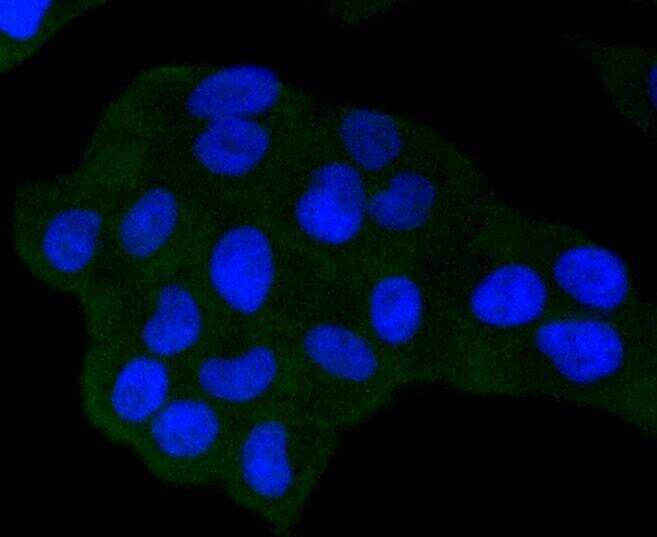
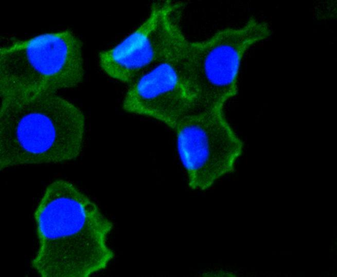
- Immunocytochemical analysis of NOX4 in A431 cells using a NOX4 Monoclonal antibody (Product # MA5-32090) as seen in green. The nuclear counter stain is DAPI (blue). Cells were fixed in paraformaldehyde, permeabilised with 0.25% Triton X100/PBS.
- Submitted by
- Invitrogen Antibodies (provider)
- Main image

- Experimental details
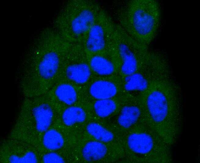
- Immunocytochemical analysis of NOX4 in A549 cells using a NOX4 Monoclonal antibody (Product # MA5-32090) as seen in green. The nuclear counter stain is DAPI (blue). Cells were fixed in paraformaldehyde, permeabilised with 0.25% Triton X100/PBS.

- Submitted by
- Invitrogen Antibodies (provider)
- Main image

- Experimental details
- Immunocytochemical analysis of NOX4 in A431 cells using a NOX4 Monoclonal antibody (Product # MA5-32090) as seen in green. The nuclear counter stain is DAPI (blue). Cells were fixed in paraformaldehyde, permeabilised with 0.25% Triton X100/PBS.
- Submitted by
- Invitrogen Antibodies (provider)
- Main image

- Experimental details
- Immunocytochemical analysis of NOX4 in A549 cells using a NOX4 Monoclonal antibody (Product # MA5-32090) as seen in green. The nuclear counter stain is DAPI (blue). Cells were fixed in paraformaldehyde, permeabilised with 0.25% Triton X100/PBS.
Supportive validation
- Submitted by
- Invitrogen Antibodies (provider)
- Main image
- Experimental details
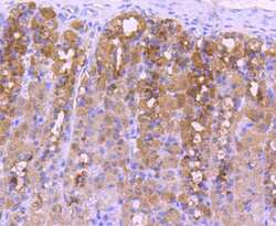
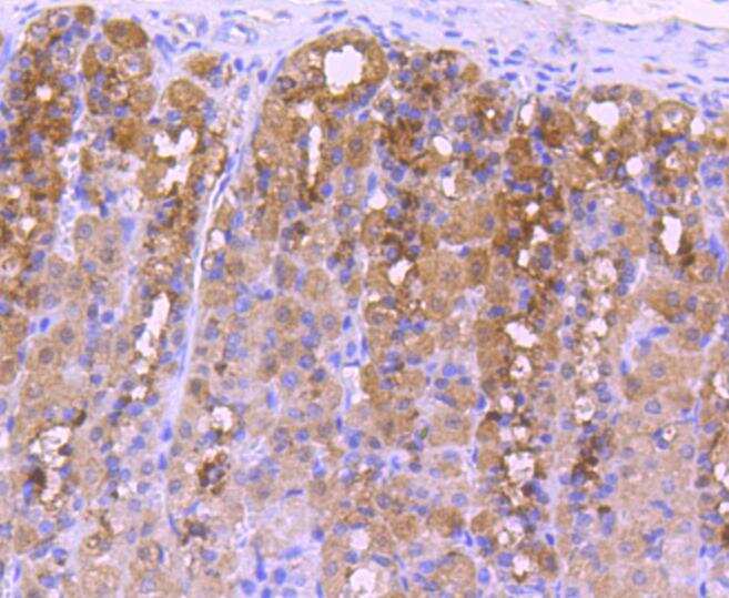
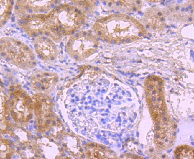
- Immunohistochemical analysis of NOX4 of paraffin-embedded rat stomach tissue using a NOX4 Monoclonal antibody (Product #MA5-32090). Counter stained with hematoxylin.
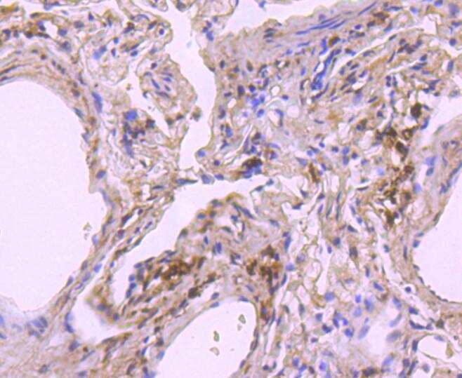
- Submitted by
- Invitrogen Antibodies (provider)
- Main image

- Experimental details
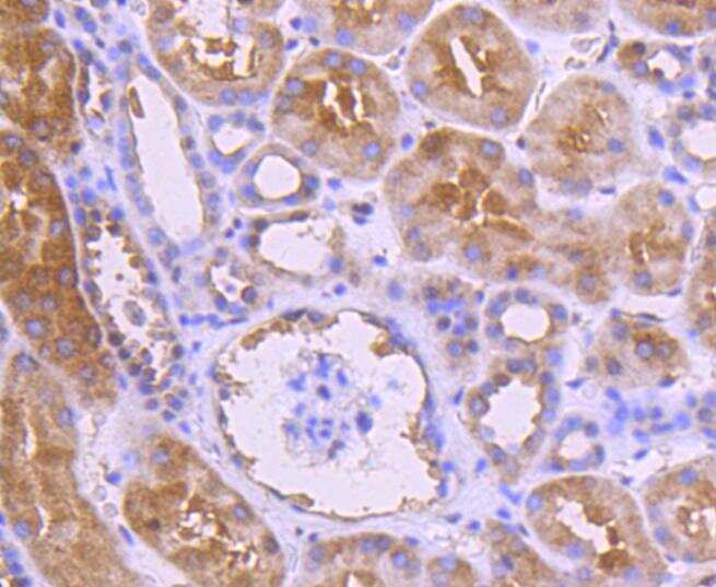
- Immunohistochemical analysis of NOX4 of paraffin-embedded Human kidney tissue using a NOX4 Monoclonal antibody (Product #MA5-32090). Counter stained with hematoxylin.
- Submitted by
- Invitrogen Antibodies (provider)
- Main image
- Experimental details
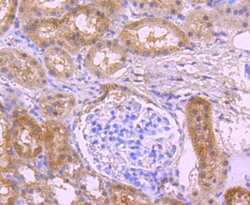
- Immunohistochemical analysis of NOX4 of paraffin-embedded rat kidney tissue using a NOX4 Monoclonal antibody (Product #MA5-32090). Counter stained with hematoxylin.
- Submitted by
- Invitrogen Antibodies (provider)
- Main image
- Experimental details
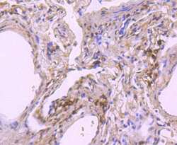
- Immunohistochemical analysis of NOX4 of paraffin-embedded Human lung tissue using a NOX4 Monoclonal antibody (Product #MA5-32090). Counter stained with hematoxylin.
Supportive validation
- Submitted by
- Invitrogen Antibodies (provider)
- Main image

- Experimental details
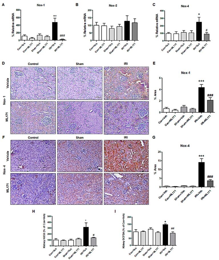
- Figure 2 Effect of ML171 on nicotinamide adenine dinucleotide phosphate (NADPH) oxidase (NOX) family subunits and oxidative stress markers in IRI. The increased mRNA expression of NOX1 and NOX4 in IRI were significantly mitigated by ML171 ( A - C ). The immunohistochemical staining of NOX1 and NOX4 showed that the increased expression of NOX1 and NOX4 in IRI was decreased by ML171 ( D - G ). The increased reactive oxygen species (ROS) including H 2 O 2 in the IRI model were significantly decreased by ML171 ( H , I ). Data represent mean +- SEM. * p < 0.05, *** p < 0.001 vs. Con + Veh, # p < 0.05, ## p < 0.01, ### p < 0.001 vs. IRI + Veh. The difference among the groups was analyzed using a one-way nonparametric ANOVA followed by Tukey's multiple comparison test.
- Submitted by
- Invitrogen Antibodies (provider)
- Main image
- Experimental details
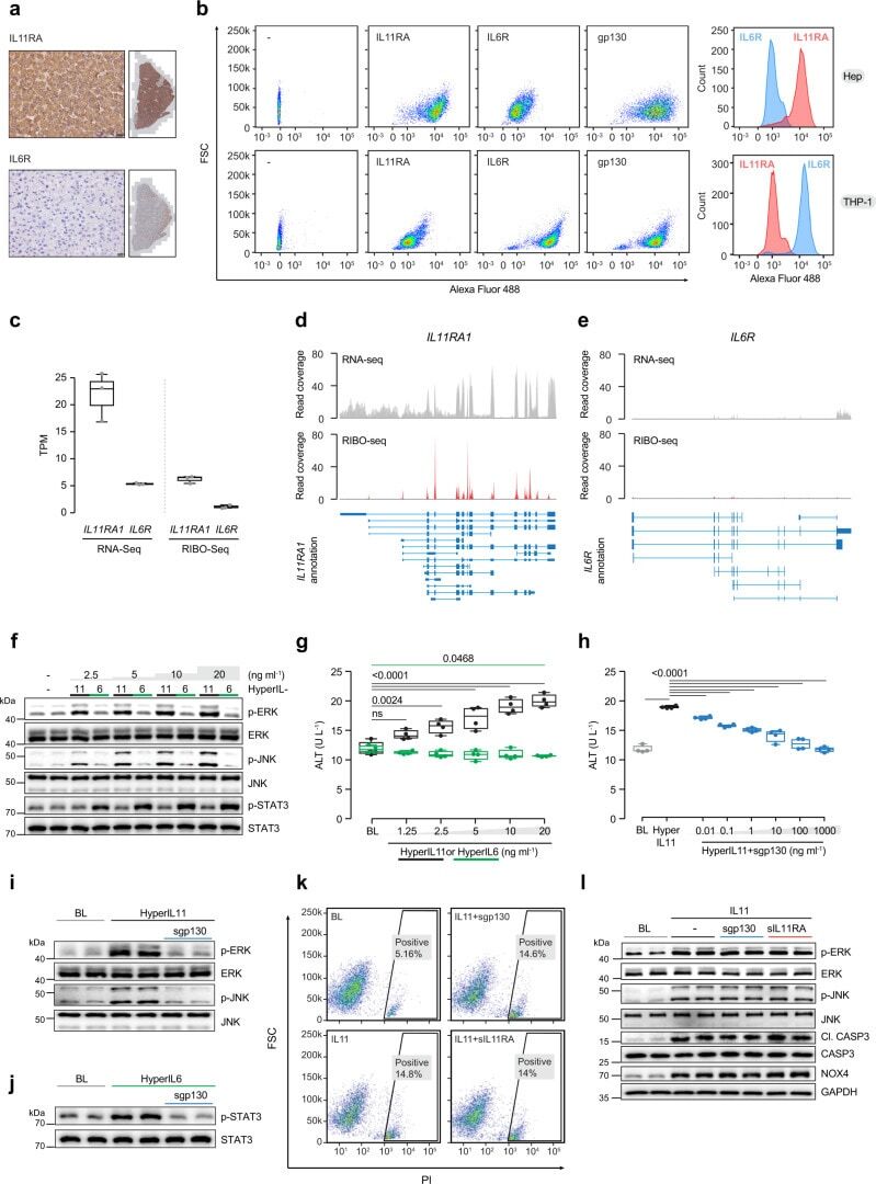
- Fig. 1 IL11RA is highly expressed in hepatocytes and IL11 cis -signaling is hepatotoxic. a Immunohistochemistry staining of IL11RA and IL6R in healthy human liver sections (scale bars, 20 um, n = 1 independent experiment, due to limited amount of human liver section). b Flow cytometry forward scatter (FSC) plots of IL11RA, IL6R, and gp130 staining and fluorescence intensity plots of IL11RA and IL6R staining on hepatocytes and THP-1. c Abundance of IL11RA1 and IL6R reads in hepatocytes at baseline based on RNA-seq (left) and Ribo-seq (right) (transcripts per million, TPM) ( n = 3). d , e Read coverage of d IL11RA1 and e IL6R transcripts based on RNA-seq (gray) and Ribo-seq (red) of primary human hepatocytes ( n = 3). f Western blots showing ERK, JNK, and STAT3 activation status and g ALT secretion ( n = 4) by hepatocytes following a dose range stimulation of either hyperIL11 or hyperIL6. h ALT levels in the supernatants of hepatocytes stimulated with hyperIL11 alone or in the presence of increasing amounts of soluble gp130 (sgp130) ( n = 4). i , j Western blots of hepatocyte lysates showing i phosphorylated ERK and JNK and their respective total expression in response to hyperIL11 stimulation alone or with sgp130 and j phospho-STAT3 and total STAT3 in response to hyperIL6 stimulation with and without sgp130. k Representative FSC plots of propidium Iodide (PI) staining of IL11-stimulated hepatocytes in the presence of sgp130 or soluble IL11RA (sIL11RA). l Western blots showing
- Submitted by
- Invitrogen Antibodies (provider)
- Main image
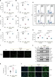
- Experimental details
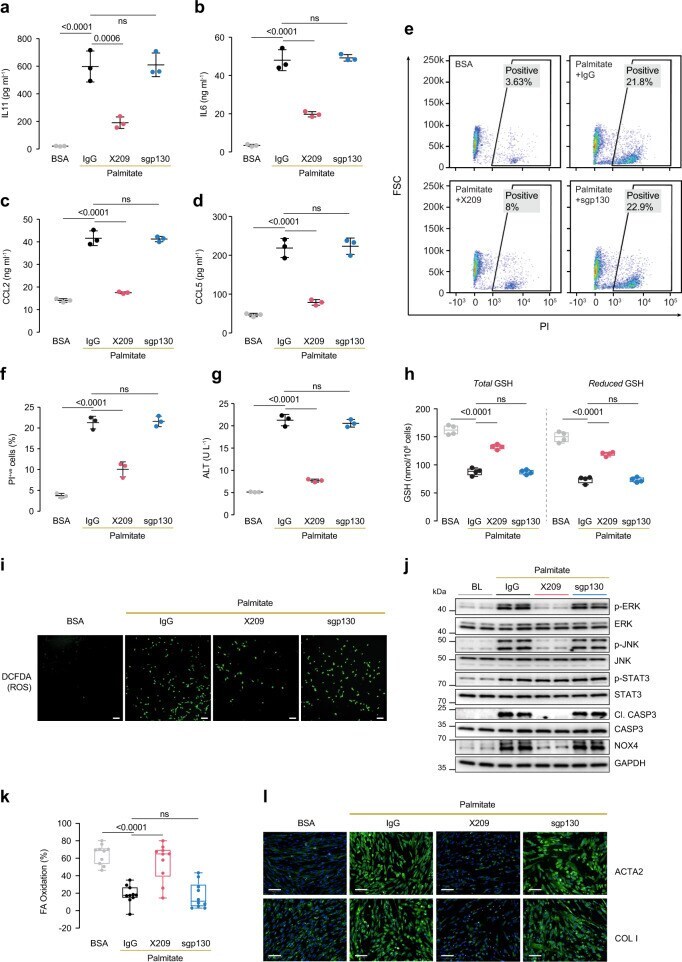
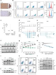
- Fig. 2 IL11 drives NASH phenotypes through autocrine effects in lipotoxic hepatocytes and paracrine activity in hepatic stellate cells. a - l Data for palmitate (0.5 mM) loading experiment on primary human hepatocytes (24 h) in the presence of either IgG (2 µg/mL), anti-IL11RA (X209, 2 µg/mL), or sgp130 (1 µg/mL). a IL11, b IL6, c CCL2, and d CCL5 protein secretion levels as measured by ELISA of supernatants ( n = 3). e Representative FSC plots and f quantification of PI +ve hepatocytes stimulated with palmitate ( n = 3). g ALT levels in supernatants ( n = 3). h Total and reduced hepatocyte glutathione (GSH) levels ( n = 4). i Representative fluorescence images of DCFDA (2',7'-dichlorofluorescein diacetate) staining for ROS detection (scale bars, 100 um) ( n = 4 independent experiments). j Western blots of phospho-ERK, ERK, phospho-JNK, JNK, cleaved caspase-3, caspase-3, NOX4, and GAPDH. Data from two independent biological experiments are shown. k Percentage of fatty acid oxidation by Seahorse assay ( n = 10). l Representative fluorescence images (scale bars, 100 um) of ACTA2 +ve cells and Collagen I immunostaining for experiment shown in Supplementary Fig. 5j ( n = 2 independent experiments, 14 measurements per condition per experiment). a - d , f , g Mean +- SD; h , k data are shown as box-and-whisker with median (middle line), 25th-75th percentiles (box), and min-max values (whiskers). a - d , f - h , k One-way ANOVA with Tukey's correction. Source data