Explore
 Validate
 Learn
Learn Western blot
Antibody data
- Antibody Data
- Antigen structure
- References [0]
- Comments [0]
- Validations
- Western blot [4]
- Immunocytochemistry [2]
- Immunohistochemistry [1]
- Other assay [1]
Submit
Validation data
Reference
Comment
Report error
- Product number
- PA5-30640 - Provider product page

- Provider
- Invitrogen Antibodies
- Product name
- TRIM25 Polyclonal Antibody
- Antibody type
- Polyclonal
- Antigen
- Recombinant protein fragment
- Description
- Recommended positive controls: A549, Jurkat, Raji, NCI-H929.
- Concentration
- 1 mg/mL
No comments: Submit comment
Supportive validation
- Submitted by
- Invitrogen Antibodies (provider)
- Main image
- Experimental details
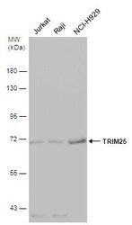
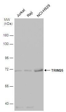
- Western Blot analysis of TRIM25 was performed by separating 30 µg of various whole cell extracts by 7.5% SDS-PAGE. Proteins were transferred to a membrane and probed with a TRIM25 Polyclonal Antibody (Product # PA5-30640) at a dilution of 1:500.
- Submitted by
- Invitrogen Antibodies (provider)
- Main image
- Experimental details
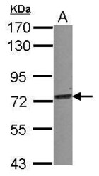
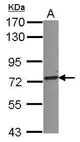
- Western Blot using TRIM25 Polyclonal Antibody (Product # PA5-30640). Sample (30 µg of whole cell lysate). A: A549 . 7.5% SDS PAGE. TRIM25 Polyclonal Antibody (Product # PA5-30640) diluted at 1:1,000.
- Submitted by
- Invitrogen Antibodies (provider)
- Main image

- Experimental details
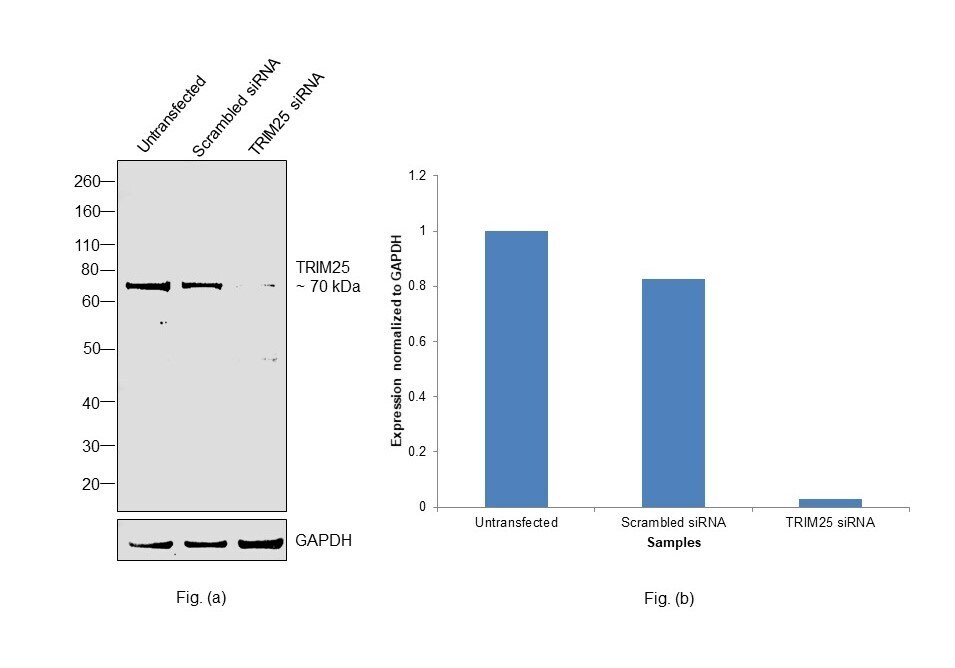
- Knockdown of E3 ubiquitin/ISG15 ligase TRIM25 was achieved by transfecting SK-BR-3 with E3 ubiquitin/ISG15 ligase TRIM25 specific siRNAs (Silencer® select Product # S15206, S15204). Western Blot analysis (Fig. a) was performed using Whole cell extracts from the E3 ubiquitin/ISG15 ligase TRIM25 knockdown cells (lane 3), non-targeting scrambled siRNA transfected cells (lane 2) and untransfected cells (lane 1). The blot was probed with TRIM25 Polyclonal Antibody (Product # PA5-30640, 1:1000 dilution) and Goat anti-Rabbit IgG (H+L) Superclonal™ Recombinant Secondary Antibody, HRP (Product # A27036, 1:20000 dilution). Densitometric analysis of this Western Blot is shown in histogram (Fig. b). Decrease in signal upon siRNA mediated knock down confirms that antibody is specific to E3 ubiquitin/ISG15 ligase TRIM25.
- Submitted by
- Invitrogen Antibodies (provider)
- Main image
- Experimental details
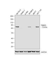
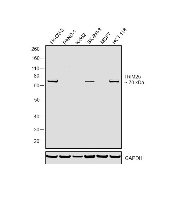
- Western Blot was performed using Anti-TRIM25 Polyclonal Antibody (Product # PA5-30640) and a 70 kDa band corresponding to E3 ubiquitin/ISG15 ligase TRIM25 was observed across tested cell lines. Whole cell extracts (40 µg lysate) of SK-O-V3 (Lane 1), PANC-1 (Lane 2), K-562 (Lane 3), SK-BR-3 (Lane 4), MCF7 (Lane 5), HCT 116 (Lane 6) were electrophoresed using NuPAGE™ 4-12% Bis-Tris Protein Gel (Product # NP0321BOX). Resolved proteins were then transferred onto a nitrocellulose membrane (Product # IB23001) by iBlot® 2 Dry Blotting System (Product # IB21001). The blot was probed with the primary antibody (1:1000 dilution) and detected by chemiluminescence with Goat anti-Rabbit IgG (H+L) Superclonal™ Recombinant Secondary Antibody, HRP (Product # A27036, 1:20000 dilution) using the iBright FL 1000 (Product # A32752). Chemiluminescent detection was performed using SuperSignal™ West Dura Extended Duration Substrate (Product # 34076).
Supportive validation
- Submitted by
- Invitrogen Antibodies (provider)
- Main image

- Experimental details
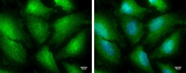
- TRIM25 Polyclonal Antibody detects TRIM25 protein at cytoplasm and nucleus by immunofluorescent analysis. Sample: HeLa cells were fixed in 4% paraformaldehyde for 10 min. Green: TRIM25 protein stained by TRIM25 Polyclonal Antibody (Product # PA5-30640) diluted at 1:100. Blue: Hoechst 33342 staining. Scale bar = 10 µm.
- Submitted by
- Invitrogen Antibodies (provider)
- Main image
- Experimental details
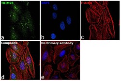
- Immunofluorescence analysis of E3 ubiquitin/ISG15 ligase TRIM25 was performed using 70% confluent log phase HeLa cells. The cells were fixed with 4% paraformaldehyde for 5 minutes, permeabilized with 0.1% Triton™ X-100 for 10 minutes, and blocked with 2% BSA for overnight at room temperature. The cells were labeled with TRIM25 Polyclonal Antibody (Product # PA5-30640) at 1:100 dilution in 0.1% BSA, incubated at 4 degree celsius overnight and then labeled with Donkey anti-Rabbit IgG (H+L) Highly Cross-Adsorbed Secondary Antibody, Alexa Fluor Plus 488 (Product # A32790), (1:2000 dilution), for 45 minutes at room temperature (Panel a: Green). Nuclei (Panel b: Blue) were stained with ProLong™ Diamond Antifade Mountant with DAPI (Product # P36962). F-actin (Panel c: Red) was stained with Rhodamine Phalloidin (Product # R415, 1:300). Panel d represents the merged image showing nucleus and cytoplasm localization. Panel e represents control cells with no primary antibody to assess background. The images were captured at 60X magnification.

Supportive validation
- Submitted by
- Invitrogen Antibodies (provider)
- Main image
- Experimental details
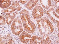
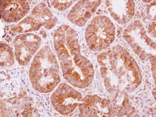
- Immunohistochemical analysis of paraffin-embedded human colon carcinoma, using TRIM25 (Product # PA5-30640) antibody at 1:500 dilution. Antigen Retrieval: EDTA based buffer, pH 8.0, 15 min.
Supportive validation
- Submitted by
- Invitrogen Antibodies (provider)
- Main image
- Experimental details
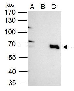
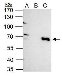
- TRIM25 antibody immunoprecipitates TRIM25 protein in IP experiments. IP Sample: HeLa whole cell lysate/extract A. 40 µg HeLa whole cell lysate/extract B. Control with 2 µg of preimmune rabbit IgG C. Immunoprecipitation of TRIM25 protein by 2 µg of TRIM25 antibody (Product # PA5-30640) 7.5% SDS-PAGE The immunoprecipitated TRIM25 protein was detected by TRIM25 antibody (Product # PA5-30640) diluted at 1:1,000.