 Explore
 Validate
 Learn
Learn Western blot
Antibody data
- Antibody Data
- Antigen structure
- References [0]
- Comments [0]
- Validations
- Western blot [1]
- Immunohistochemistry [3]
Submit
Validation data
Reference
Comment
Report error
- Product number
- TA328875 - Provider product page

- Provider
- OriGene
- Product name
- Rabbit Polyclonal Anti-Melatonin Receptor type 2
- Antibody type
- Polyclonal
- Description
- Rabbit Polyclonal Anti-Melatonin Receptor type 2
- Host
- Rabbit
- Conjugate
- Unconjugated
- Epitope
- Mtnr1b
- Antibody clone number
- NULL
- Vial size
- 50 µl
- Concentration
- NULL
No comments: Submit comment
Supportive validation
- Submitted by
- OriGene (provider)
- Main image

- Experimental details
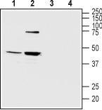
- Western blot analysis of mouse (lanes 1 and 3) and rat (lanes 2 and 4) brain lysates: 1,2. Anti-Melatonin Receptor Type 2 antibody, (1:200). 3,4. Anti-Melatonin Receptor Type 2 antibody, preincubated with the control peptide antigen.
- Validation comment
- WB
Supportive validation
- Submitted by
- OriGene (provider)
- Main image
- Experimental details
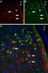
- IHC staining of rat paraventricular nucleus sections using Anti-Melatonin Receptor Type 2 antibody , (1:600) and guinea pig Anti-NaV1.2 antibody, (1:2000). A. Melatonin Receptor Type 2 staining (red) (arrows). B. The same section labeled for NaV1.2 (green). C. Merge of A and B demonstrates partial co-localization of Melatonin Receptor Type 2 and NaV1.2 in the paraventricular nucleus (PVN). For orientation, note location with respect to the third ventricle (3rd V).
- Validation comment
- IHC
- Submitted by
- OriGene (provider)
- Main image
- Experimental details
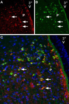
- IHC staining of perfusion-fixed frozen brain sections using Anti-Melatonin Receptor Type 2 antibody, (1:600) and Anti-Angiotensin II Receptor Type-2 (extracellular)-ATTO-488, (1:100). A. Melatonin Receptor Type 2 staining (red) (arrows). B. The same section labeled for Angiotensin II Receptor Type-2 (green). C. Merge of the two images suggests considerable co-localization in the paraventricular nucleus (arrows). For orientation, note localization with respect to 3rd ventricle (3rd V).
- Validation comment
- IHC

- Submitted by
- OriGene (provider)
- Main image
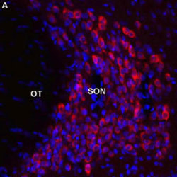
- Experimental details
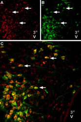
- Expression of Melatonin receptor type 2 in rat supraoptic nucleus. Immunohistochemical staining of perfusion-fixed frozen brain sections using Anti-Melatonin Receptor Type 2 antibody, (1:600), (red). Melatonin Receptor Type 2 is expressed discretely in the supraoptic nucleus (SON), adjacent to the optric tract (OT). DAPI is used as the counterstain (blue).
- Validation comment
- IHC
