Explore
 Validate
 Learn
Learn Western blot
 ELISA
ELISAAntibody data
- Antibody Data
- Antigen structure
- References [0]
- Comments [0]
- Validations
- Western blot [1]
- Immunohistochemistry [1]
- Flow cytometry [1]
Submit
Validation data
Reference
Comment
Report error
- Product number
- NBP1-97941-100ul - Provider product page

- Provider
- Novus Biologicals
- Product name
- Mouse Monoclonal PNPO Antibody
- Antibody type
- Monoclonal
- Description
- Protein G purified.
- Reactivity
- Human
- Host
- Mouse
- Isotype
- IgG
- Vial size
- 100 ul
- Concentration
- 1 mg/ml
- Storage
- Store at 4C short term. Aliquot and store at -20C long term. Avoid freeze-thaw cycles.
No comments: Submit comment
Supportive validation
- Submitted by
- Novus Biologicals (provider)
- Main image
- Experimental details
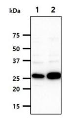
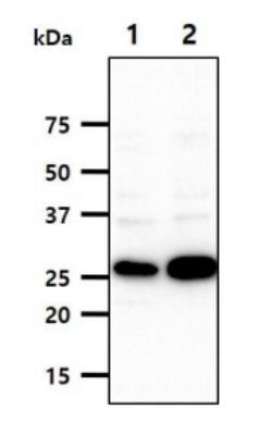
- Western Blot: PNPO Antibody (2C7) [NBP1-97941] - The Cell lysates (40ug) were resolved by SDS-PAGE, transferred to PVDF membrane and probed with anti-human PNPO antibody (1:1000). Proteins were visualized using a goat anti-mouse secondary antibody conjugated to HRP and an ECL detection system. Lane 1.: A549 cell lysate Lane 2.: HepG2 cell lysate
Supportive validation
- Submitted by
- Novus Biologicals (provider)
- Main image
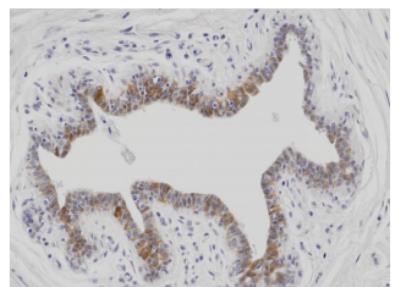
- Experimental details
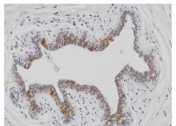
- Immunohistochemistry-Paraffin: PNPO Antibody (2C7) [NBP1-97941] - Paraffin embedded sections of human breast cancer tissue were incubated with anti-human PNPO (1:150) for 2 hours at room temperature. Antigen retrieval was performed in 0.1M sodium citrate buffer and detected using Diaminobenzidine(DAB).
Supportive validation
- Submitted by
- Novus Biologicals (provider)
- Main image
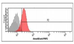
- Experimental details
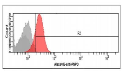
- Flow Cytometry: PNPO Antibody (2C7) [NBP1-97941] - Flow cytometry analysis of PNPO in Jurkat cells. The cell was stained at 2-5ug for 1x10⁶cells (red). A Goat anti mouse IgG (Alexa fluor 488) was used as the secondary antibody. Mouse monoclonal IgG was used as the isotype control (dark gray), cells without incubation with primary and secondary antibody was used as the negative control (light gray).