 Explore
 Validate
 Learn
Learn Western blot
 ELISA
ELISAAntibody data
- Antibody Data
- Antigen structure
- References [0]
- Comments [0]
- Validations
- Western blot [2]
- Immunohistochemistry [2]
Submit
Validation data
Reference
Comment
Report error
- Product number
- LS-B10629 - Provider product page

- Provider
- LSBio
- Product name
- IHC-plus™ PFDN5 / MM1 Antibody LS-B10629
- Antibody type
- Polyclonal
- Description
- Caprylic acid and ammonium sulfate precipitation
- Reactivity
- Human
- Host
- Rabbit
- Isotype
- IgG
- Storage
- Aliquot and store at -20°C or -80°C. Avoid freeze-thaw cycles.
No comments: Submit comment
Enhanced validation
- Submitted by
- LSBio (provider)
- Enhanced method
- Genetic validation
- Main image
- Experimental details
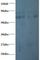
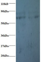
- Western blot of Prefoldin subunit 5 antibody at 2 ug/ml. Lane 1: EC109whole cell lysate. Lane 2: 293T whole cell lysate. Secondary: Goat polyclonal to Rabbit IgG at 1:15000 dilution. Predicted band size: 17 kDa. Observed band size: 70 kDa Additional bands at: 80 kDa. We are unsure as to the identity of these extra band. This image was taken for the unconjugated form of this product. Other forms have not been tested.
- Submitted by
- LSBio (provider)
- Enhanced method
- Genetic validation
- Main image
- Experimental details
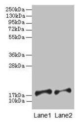
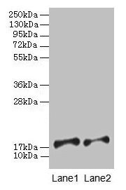
- Western blot All lanes: Prefoldin subunit 5 antibody at 2µg/ml Lane 1: EC109whole cell lysate Lane 2: 293T whole cell lysate Secondary Goat polyclonal to rabbit IgG at 1/15000 dilution Predicted band size: 18, 8, 13 kDa Observed band size: 18 kDa
Supportive validation
- Submitted by
- LSBio (provider)
- Enhanced method
- Genetic validation
- Main image
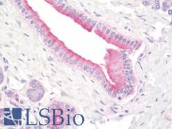
- Experimental details
- Anti-PFDN5 / MM1 antibody IHC staining of human pancreas. Immunohistochemistry of formalin-fixed, paraffin-embedded tissue after heat-induced antigen retrieval. Antibody concentration 10 ug/ml. This image was taken for the unconjugated form of this product. Other forms have not been tested.
- Submitted by
- LSBio (provider)
- Enhanced method
- Genetic validation
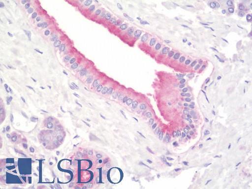
- Main image

- Experimental details
- Anti-PFDN5 / MM1 antibody IHC staining of human pancreas. Immunohistochemistry of formalin-fixed, paraffin-embedded tissue after heat-induced antigen retrieval. Antibody concentration 10 ug/ml. This image was taken for the unconjugated form of this product. Other forms have not been tested.
