Explore
 Validate
 Learn
Learn Western blot
 Immunohistochemistry
Antibody data
- Antibody Data
- Antigen structure
- References [1]
- Comments [0]
- Validations
- Western blot [1]
- Immunocytochemistry [1]
Submit
Validation data
Reference
Comment
Report error
- Product number
- HPA035015 - Provider product page

- Provider
- Atlas Antibodies
- Proper citation
- Atlas Antibodies Cat#HPA035015, RRID:AB_10603117
- Product name
- Anti-STAG1
- Antibody type
- Polyclonal
- Description
- Polyclonal Antibody against Human STAG1, Gene description: stromal antigen 1, Alternative Gene Names: SA-1, SCC3A, Validated applications: WB, IHC, Uniprot ID: Q8WVM7, Storage: Store at +4°C for short term storage. Long time storage is recommended at -20°C.
- Reactivity
- Human, Mouse, Rat
- Host
- Rabbit
- Conjugate
- Unconjugated
- Isotype
- IgG
- Vial size
- 100 µl
- Concentration
- 0.1 mg/ml
- Storage
- Store at +4°C for short term storage. Long time storage is recommended at -20°C.
- Handling
- The antibody solution should be gently mixed before use.
Submitted references Synthetic lethal interaction between the tumour suppressor STAG2 and its paralog STAG1
Benedetti L, Cereda M, Monteverde L, Desai N, Ciccarelli F
Oncotarget 2017;8(23):37619-37632
Oncotarget 2017;8(23):37619-37632
No comments: Submit comment
Enhanced validation
- Submitted by
- Atlas Antibodies (provider)
- Enhanced method
- Genetic validation
- Main image

- Experimental details
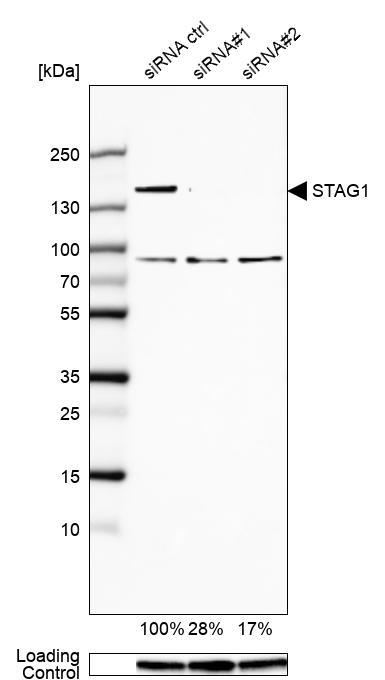
- Western blot analysis in U-138MG cells transfected with control siRNA, target specific siRNA probe #1 and #2, using Anti-STAG1 antibody. Remaining relative intensity is presented. Loading control: Anti-GAPDH.
- Sample type
- Human
- Protocol
- Protocol
Enhanced validation
- Submitted by
- 55af80e3e0991
- Enhanced method
- Genetic validation
- Main image
- Experimental details
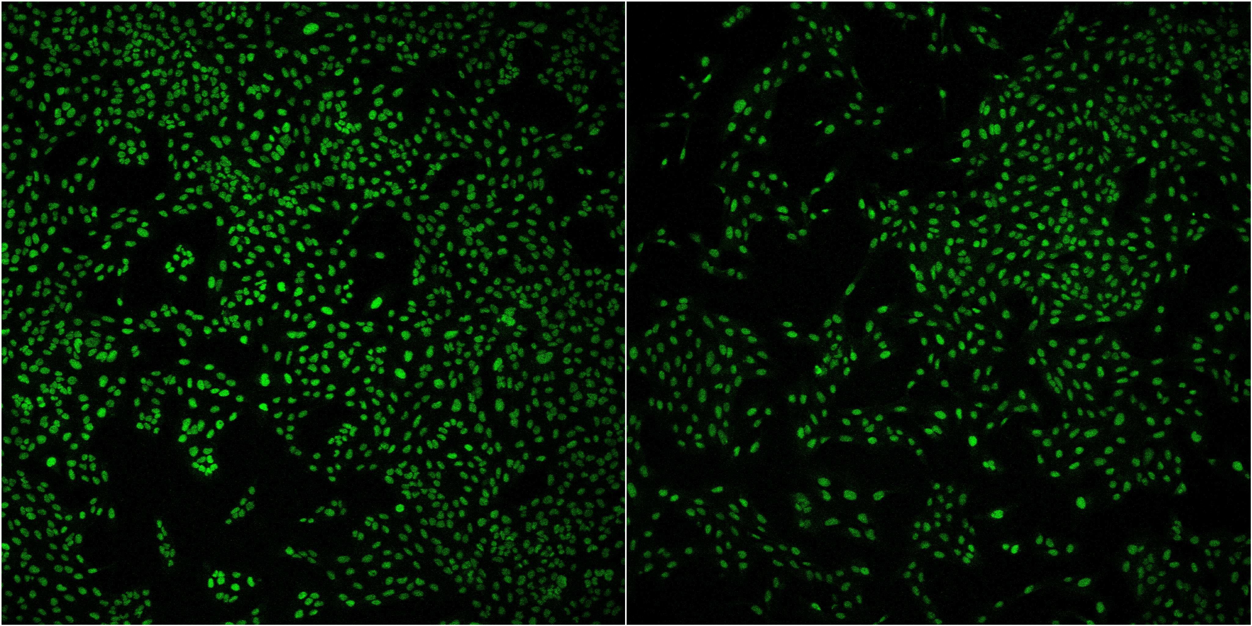
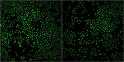
- Confocal images of immunofluorescently stained human U-2 OS cells.The protein STAG1 is shown in green. The image to the left show cells transfected with control siRNA and the image to the right show cells where STAG1 has been downregulated with specific siRNA.
- Sample type
- U-2 OS cells
- Primary Ab dilution
- 1:34
- Secondary Ab
- Secondary Ab
- Secondary Ab dilution
- 1:800
- Knockdown/Genetic Approaches Application
- Immunocytochemistry