 Explore
 Validate
 Learn
Learn Western blot
Antibody data
- Antibody Data
- Antigen structure
- References [0]
- Comments [0]
- Validations
- Western blot [4]
- Immunocytochemistry [3]
- Immunohistochemistry [1]
Submit
Validation data
Reference
Comment
Report error
- Product number
- PA5-57115 - Provider product page

- Provider
- Invitrogen Antibodies
- Product name
- STAG1 Polyclonal Antibody
- Antibody type
- Polyclonal
- Antigen
- Recombinant full-length protein
- Description
- Immunogen sequence: TSELPVLQDS TNETTAHSDA GSELEETEVK GKRKRGRPGR PPSTNKKPRK SPGEKSR Highest antigen sequence identity to the following orthologs: Mouse - 100%, Rat - 100%.
- Reactivity
- Human, Mouse, Rat
- Host
- Rabbit
- Isotype
- IgG
- Vial size
- 100 µL
- Concentration
- 0.1 mg/mL
- Storage
- Store at 4°C short term. For long term storage, store at -20°C, avoiding freeze/thaw cycles.
No comments: Submit comment
Supportive validation
- Submitted by
- Invitrogen Antibodies (provider)
- Main image

- Experimental details
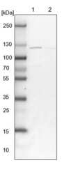
- Western blot analysis of STAG1 in Lane 1: Marker (kDa) 230, 130, 95, 72, 56, 36, 28, 17, 11; Lane 2: Human cell line RT-4. Samples were probed using a STAG1 Polyclonal Antibody (Product # PA5-57115).

- Submitted by
- Invitrogen Antibodies (provider)
- Main image
- Experimental details
- Western blot analysis of STAG1 in Lane 1: NIH-3T3 cell lysate (Mouse embryonic fibroblast cells); Lane 2: NBT-II cell lysate (Rat Wistar bladder tumour cells). Samples were probed using a STAG1 Polyclonal Antibody (Product # PA5-57115).

- Submitted by
- Invitrogen Antibodies (provider)
- Main image

- Experimental details
- Knockdown of Cohesin subunit SA-1 was achieved by transfecting MCF 10A with Cohesin subunit SA-1 specific siRNAs (Silencer® select Product # s20075, s20076). Western blot analysis (Fig. a) was performed using Whole cell extracts from the Cohesin subunit SA-1 knockdown cells (lane 3), non-targeting scrambled siRNA transfected cells (lane 2) and untransfected cells (lane 1). The blot was probed with STAG1 Polyclonal Antibody (Product # PA5-57115, 1:3000 ) and Goat anti-Rabbit IgG (H+L) Superclonal™ Recombinant Secondary Antibody, HRP (Product # A27036, 1:20,000). Densitometric analysis of this western blot is shown in histogram (Fig. b). Decrease in signal upon siRNA mediated knock down confirms that antibody is specific to Cohesin subunit SA-1.

- Submitted by
- Invitrogen Antibodies (provider)
- Main image

- Experimental details
- Western blot was performed using Anti-STAG1 Polyclonal Antibody (Product # PA5-57115) and a 150kDa band corresponding to Cohesin subunit SA-1 was observed. Whole cell extracts (30 µg lysate) of HeLa (Lane 1), Jurkat (Lane 2), MCF7 (Lane 3) and PC-12 (Lane 4) were electrophoresed using NuPAGE™ 4-12% Bis-Tris Protein Gel (Product # NP0322BOX). Resolved proteins were then transferred onto a nitrocellulose membrane (Product # IB23001) by iBlot® 2 Dry Blotting System (Product # IB21001). The blot was probed with the primary antibody (1:3000) and detected by chemiluminescence with Goat anti-Rabbit IgG (H+L) Superclonal™ Recombinant Secondary Antibody, HRP (Product # A27036,1:20,000 using the iBright™ FL1500 Imaging System (Product # A44115). Chemiluminescent detection was performed using SuperSignal™ West Pico PLUS Chemiluminescent Substrate (Product # 34580).

Supportive validation
- Submitted by
- Invitrogen Antibodies (provider)
- Main image

- Experimental details
- Immunofluorescent staining of STAG1 in human cell line U-251 MG shows positivity in nucleus but excluded from the nucleoli. Samples were probed using a STAG1 Polyclonal Antibody (Product # PA5-57115).

- Submitted by
- Invitrogen Antibodies (provider)
- Main image

- Experimental details
- Immunofluorescent staining of STAG1 in human cell line A-431 using a STAG1 Polyclonal Antibody (Product # PA5-57115) shows localization to nucleus and nuclear bodies.

- Submitted by
- Invitrogen Antibodies (provider)
- Main image
- Experimental details
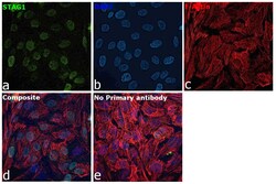
- Immunofluorescence analysis of Cohesin subunit SA-1 was performed using 70% confluent log phase MCF 10A cells. The cells were fixed with 4% paraformaldehyde for 10 minutes, permeabilized with 0.1% Triton™ X-100 for 15 minutes, and blocked with 2% BSA for 1 hour at room temperature. The cells were labeled with STAG1 Polyclonal Antibody (Product # PA5-57115) at 0.5 ug/ml in 0.1% BSA, incubated at 4 degree celsius overnight and then labeled with Donkey anti-Rabbit IgG (H+L) Highly Cross-Adsorbed Secondary Antibody, Alexa Fluor Plus 488 (Product # A32790), (1:2000), for 45 minutes at room temperature (Panel a: Green). Nuclei (Panel b:Blue) were stained with ProLong™ Diamond Antifade Mountant with DAPI (Product # P36962). F-actin (Panel c: Red) was stained with Rhodamine Phalloidin (Product # R415, 1:300). Panel d represents the merged image showing nuclear localization. Panel e represents control cells with no primary antibody to assess background. The images were captured at 60X magnification.

Supportive validation
- Submitted by
- Invitrogen Antibodies (provider)
- Main image

- Experimental details
- Immunohistochemical staining of STAG1 in human breast tissue shows strong nuclear positivity in glandular cells. Samples were probed using a STAG1 Polyclonal Antibody (Product # PA5-57115).
