 Explore
 Validate
 Learn
Learn Western blot
Antibody data
- Antibody Data
- Antigen structure
- References [0]
- Comments [0]
- Validations
- Western blot [6]
- Immunocytochemistry [4]
- Immunoprecipitation [1]
- Other assay [1]
Submit
Validation data
Reference
Comment
Report error
- Product number
- MA5-31524 - Provider product page

- Provider
- Invitrogen Antibodies
- Product name
- STAG1 Monoclonal Antibody (GT1515)
- Antibody type
- Monoclonal
- Antigen
- Synthetic peptide
- Description
- Keep as concentrated solution. Predicted reactivity: Mouse (100%), Rat (100%), Xenopus laevis (100%). Positive Control: 293T, A431, HeLa, HepG2. Store product as a concentrated solution. Centrifuge briefly prior to opening the vial.
- Reactivity
- Human, Rat
- Host
- Mouse
- Isotype
- IgG
- Antibody clone number
- GT1515
- Vial size
- 100 μL
- Concentration
- 0.9 mg/mL
- Storage
- Store at 4°C short term. For long term storage, store at -20°C, avoiding freeze/thaw cycles.
No comments: Submit comment
Supportive validation
- Submitted by
- Invitrogen Antibodies (provider)
- Main image
- Experimental details
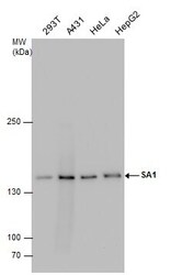
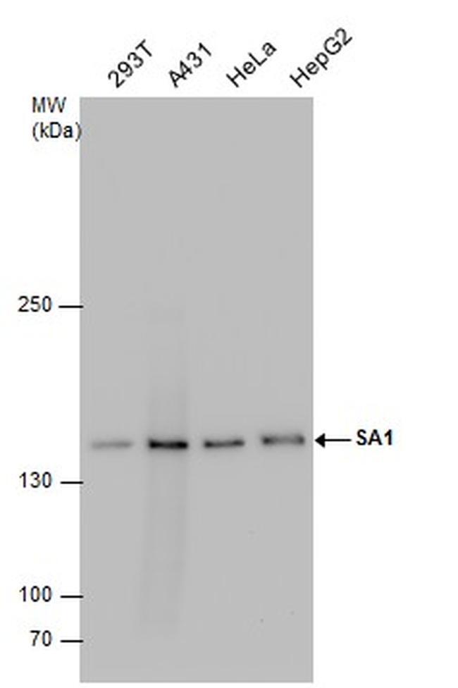
- Western Blot using STAG1 Monoclonal Antibody (GT1515) (Product # MA5-31524). Various whole cell extracts (30 µg) were separated by 5% SDS-PAGE, and the membrane was blotted with STAG1 Polyclonal Antibody STAG1 Monoclonal Antibody (GT1515) (Product # MA5-31524) diluted at 1:1,000.
- Submitted by
- Invitrogen Antibodies (provider)
- Main image
- Experimental details
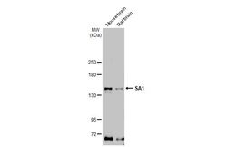
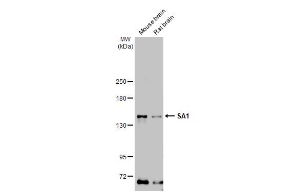
- Western Blot using STAG1 Monoclonal Antibody (GT1515) (Product # MA5-31524). Various tissue extracts (50 µg) were separated by 5% SDS-PAGE, and the membrane was blotted with STAG1 Monoclonal Antibody (GT1515) (Product # MA5-31524) diluted at 1:1,000. The HRP-conjugated anti-mouse IgG antibody was used to detect the primary antibody.
- Submitted by
- Invitrogen Antibodies (provider)
- Main image
- Experimental details
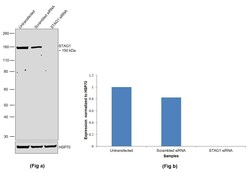
- Knockdown of Cohesin subunit SA-1 was achieved by transfecting MCF 10A with Cohesin subunit SA-1 specific siRNAs (Silencer® select Product # s20075, s20076). Western blot analysis (Fig. a) was performed using Whole cell extracts from the Cohesin subunit SA-1 knockdown cells (lane 3), non-targeting scrambled siRNA transfected cells (lane 2) and untransfected cells (lane 1). The blot was probed with STAG1 Monoclonal Antibody (GT1515) (Product # MA5-31524, 1:1000 ) and Goat anti-Mouse IgG (H+L) Superclonal™ Recombinant Secondary Antibody, HRP (Product # A28177, 1:20,000). Densitometric analysis of this western blot is shown in histogram (Fig. b). Decrease in signal upon siRNA mediated knock down confirms that antibody is specific to Cohesin subunit SA-1.

- Submitted by
- Invitrogen Antibodies (provider)
- Main image
- Experimental details
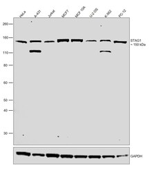
- Western blot was performed using Anti-STAG1 Monoclonal Antibody (GT1515) (Product # MA5-31524) and a 150 kDa band corresponding to Cohesin subunit SA-1 was observed. Nuclear enriched extracts (30 µg lysate) of HeLa (Lane 1), A-431 (Lane 2), Jurkat (Lane 3), MCF7 (Lane 4), MCF 10A (Lane 5), U-2 OS (Lane 6), K-562 (Lane 7) and PC-12 (Lane 8) were electrophoresed using NuPAGE™ 4-12% Bis-Tris Protein Gel (Product # NP0322BOX). Resolved proteins were then transferred onto a nitrocellulose membrane (Product # IB23001) by iBlot® 2 Dry Blotting System (Product # IB21001). The blot was probed with the primary antibody (1:1000) and detected by chemiluminescence with Goat anti-Mouse IgG (H+L) Superclonal™ Recombinant Secondary Antibody, HRP (Product # A28177,1:20,000) using the iBright FL 1000 (Product # A32752). Chemiluminescent detection was performed using SuperSignal™ West Dura Extended Duration Substrate (Product # 34076).
- Submitted by
- Invitrogen Antibodies (provider)
- Main image
- Experimental details
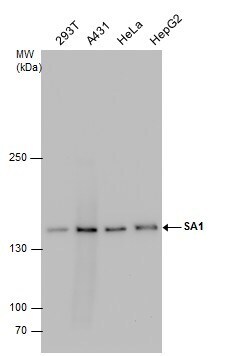
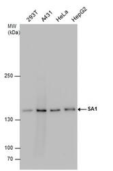
- Western blot analysis of STAG1 in various whole cell extracts using STAG1 monoclonal antibody (Product # MA5-31524) using 30 µg of sample at a dilution of 1:1000. Prior to incubation with primary antibody, the sample was separated on 5% SDS-PAGE.
- Submitted by
- Invitrogen Antibodies (provider)
- Main image
- Experimental details
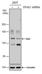
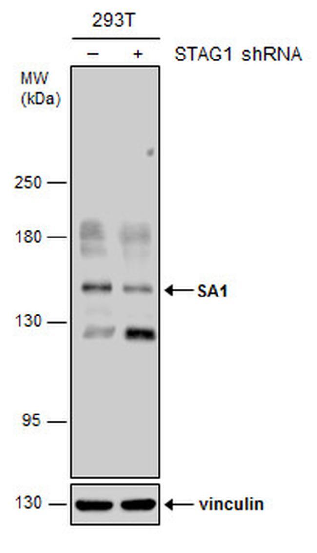
- Western blot analysis of STAG1 in non-transfected (–) and transfected (+) 293T whole cell extracts using STAG1 monoclonal antibody (Product # MA5-31524) using 30 µg of sample at a dilution of 1:500. Prior to incubation with primary antibody, the sample was separated on 5% SDS-PAGE.
Supportive validation
- Submitted by
- Invitrogen Antibodies (provider)
- Main image
- Experimental details
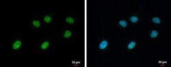
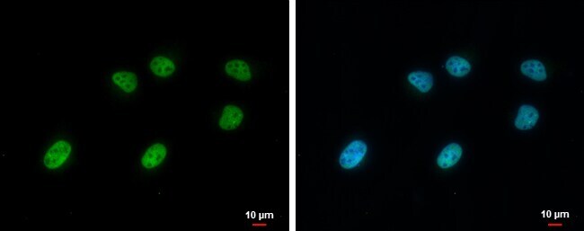
- STAG1 Monoclonal Antibody (GT1515) detects SA1 protein at nucleus by immunofluorescent analysis. Sample: HeLa cells were fixed in 4% paraformaldehyde at RT for 15 min. Green: SA1 protein stained by STAG1 Monoclonal Antibody (GT1515) (Product # MA5-31524) diluted at 1:1,000. Blue: Hoechst 33342 staining. Scale bar = 10 µm.

- Submitted by
- Invitrogen Antibodies (provider)
- Main image
- Experimental details
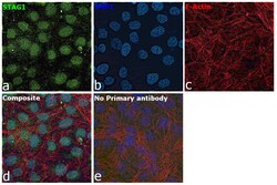
- Immunofluorescence analysis of Cohesin subunit SA-1 was performed using 70% confluent log phase MCF 10A cells. The cells were fixed with 4% paraformaldehyde for 10 minutes, permeabilized with 0.1% Triton™ X-100 for 15 minutes, and blocked with 2% BSA for 1 hour at room temperature. The cells were labeled with STAG1 Monoclonal Antibody (GT1515) (Product # MA5-31524) at 1:500 in 0.1% BSA, incubated at 4 degree celsius overnight and then labeled with Donkey anti-Mouse IgG (H+L) Highly Cross-Adsorbed Secondary Antibody, Alexa Fluor Plus 488 (Product # A32766), (1:2000), for 45 minutes at room temperature (Panel a: Green). Nuclei (Panel b:Blue) were stained with ProLong™ Diamond Antifade Mountant with DAPI (Product # P36962). F-actin (Panel c: Red) was stained with Rhodamine Phalloidin (Product # R415, 1:300). Panel d represents the merged image showing predominantly nuclear localization. Panel e represents control cells with no primary antibody to assess background. The images were captured at 60X magnification.

- Submitted by
- Invitrogen Antibodies (provider)
- Main image

- Experimental details
- Immunocytochemistry analysis of STAG1 in 4% paraformaldehyde-fixed HeLa cells using STAG1 monoclonal antibody (Product # MA5-31524) at a dilution of 1:1000. Sample was then incubated with Hoechst secondary antibody.
- Submitted by
- Invitrogen Antibodies (provider)
- Main image

- Experimental details
- Immunofluorescence analysis of Cohesin subunit SA-1 was performed using 70% confluent log phase MCF 10A cells. The cells were fixed with 4% paraformaldehyde for 10 minutes, permeabilized with 0.1% Triton™ X-100 for 15 minutes, and blocked with 2% BSA for 1 hour at room temperature. The cells were labeled with STAG1 Monoclonal Antibody (GT1515) (Product # MA5-31524) at 1:500 in 0.1% BSA, incubated at 4 degree celsius overnight and then labeled with Donkey anti-Mouse IgG (H+L) Highly Cross-Adsorbed Secondary Antibody, Alexa Fluor Plus 488 (Product # A32766), (1:2000), for 45 minutes at room temperature (Panel a: Green). Nuclei (Panel b:Blue) were stained with ProLong™ Diamond Antifade Mountant with DAPI (Product # P36962). F-actin (Panel c: Red) was stained with Rhodamine Phalloidin (Product # R415, 1:300). Panel d represents the merged image showing predominantly nuclear localization. Panel e represents control cells with no primary antibody to assess background. The images were captured at 60X magnification.

Supportive validation
- Submitted by
- Invitrogen Antibodies (provider)
- Main image

- Experimental details
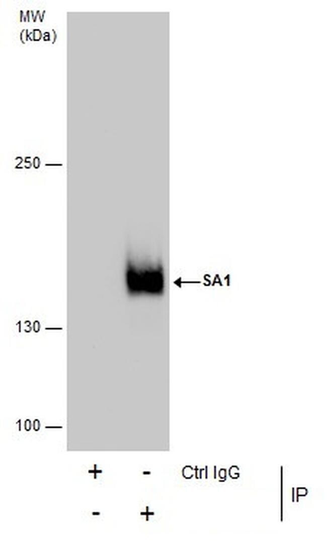
- Immunoprecipitation analysis of STAG1 in HeLa whole cell extracts with STAG1 monoclonal antibody (Product # MA5-31524) using 5 µg of sample. Sample was then incubated with anti-mouse IgG secondary antibody.
Supportive validation
- Submitted by
- Invitrogen Antibodies (provider)
- Main image
- Experimental details
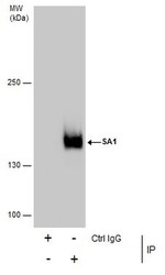
- Immunoprecipitation analysis of STAG1 in HeLa whole cell extracts with STAG1 monoclonal antibody (Product # MA5-31524) using 5 µg of sample. Sample was then incubated with anti-mouse IgG secondary antibody.
