 Explore
 Validate
 Learn
LearnMA5-37991
antibody from Invitrogen Antibodies
Targeting: TNPO3
IPO12, LGMD1F, MTR10A, TRN-SR, TRN-SR2
 Western blot
 ELISA
ELISAAntibody data
- Antibody Data
- Antigen structure
- References [0]
- Comments [0]
- Validations
- Western blot [2]
- Immunocytochemistry [5]
- Immunohistochemistry [2]
Submit
Validation data
Reference
Comment
Report error
- Product number
- MA5-37991 - Provider product page

- Provider
- Invitrogen Antibodies
- Product name
- TNPO3 Recombinant Rabbit Monoclonal Antibody (0U3Q1)
- Antibody type
- Monoclonal
- Antigen
- Synthetic peptide
- Description
- Positive Samples: HeLa, RD, Jurkat, Mouse lung, Mouse testis, Mouse thymus, Rat testis Immunogen sequence: QVMNQLGQQL VSQLLHTCCF CLPPYTLPDV AEVLWEIMQV DRPTFCRWLE NSLKGLPKET TVGAVTVTHK QLTDFHKQVT SAEECKQVCW ALRDFTRLFR
- Reactivity
- Human, Mouse, Rat
- Host
- Rabbit
- Isotype
- IgG
- Antibody clone number
- 0U3Q1
- Vial size
- 100 μL
- Concentration
- 1.65 mg/mL
- Storage
- -20°C, Avoid Freeze/Thaw Cycles
No comments: Submit comment
Supportive validation
- Submitted by
- Invitrogen Antibodies (provider)
- Main image
- Experimental details
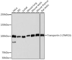
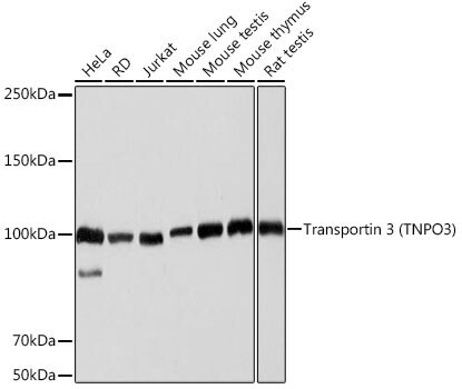
- Western blot analysis of TNPO3 in extracts of various cell lines. Samples were incubated with TNPO3 Monoclonal antibody (Product # MA5-37991) using a dilution of 1:1,000, followed by HRP Goat Anti-Rabbit IgG (H+L) at a dilution of 1:10,000. Lysates/proteins: 25 µg per lane. Blocking buffer: 3% nonfat dry milk in TBST. Detection: ECL Basic Kit. Exposure time: 3s.
- Submitted by
- Invitrogen Antibodies (provider)
- Main image
- Experimental details
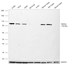
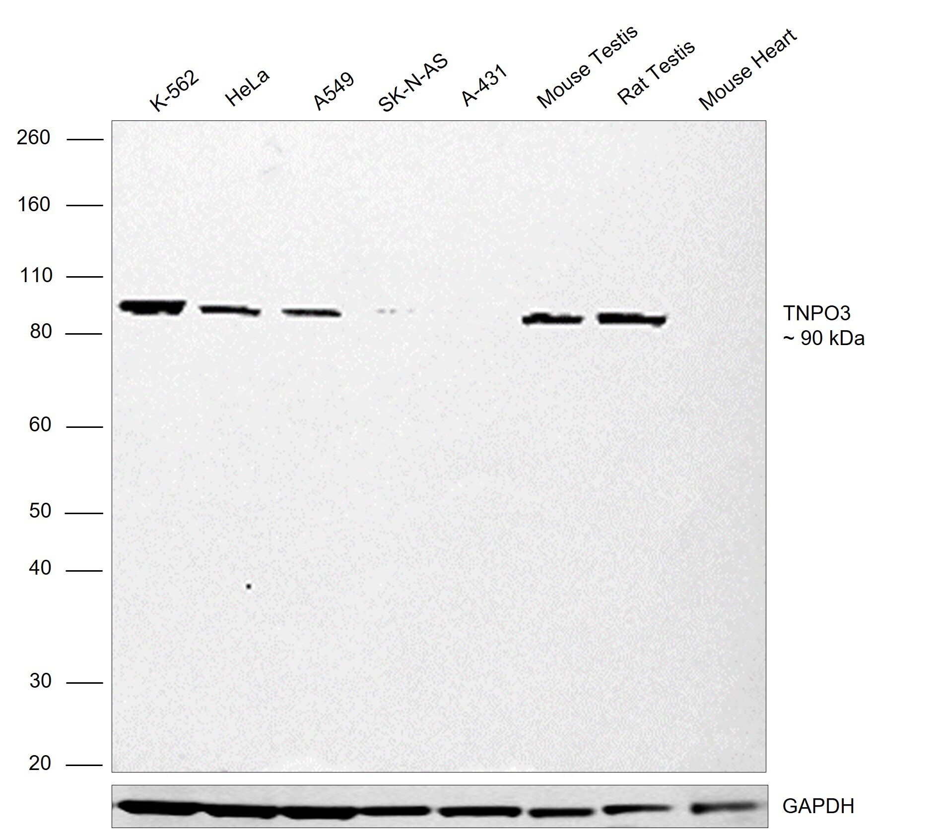
- Western blot was performed using TNPO3 Recombinant Rabbit Monoclonal Antibody (ARC2310) (Product # MA5-37991) and a 90 kDa band corresponding to TNPO3 was observed across K-562, HeLa, A549, Mouse testis and Rat testis which are high expression models and no band was seen in SK-N-AS, A-431 and Mouse heart which are low expression models. Whole cell extracts (30 µg lysate) of K-562 (Lane 1), HeLa (Lane 2), A549 (Lane 3), SK-N-AS (Lane 4), A-431 (Lane 5), Mouse Testis (Lane 6), Rat Testis (Lane 7), Mouse Heart (Lane 8) were electrophoresed using NuPAGE™ 4-12% Bis-Tris Protein Gel (Product # NP0321BOX), 10 well. Resolved proteins were then transferred onto a nitrocellulose membrane (Product # IB23001) by iBlot® 2 Dry Blotting System (Product # IB21001). The blot was probed with the primary antibody (1:1,000) and detected by chemiluminescence with Goat anti-Rabbit IgG (H+L) Superclonal™ Recombinant Secondary Antibody, HRP (Product # A27036, 1:10,000) using the iBright™ FL1500 Imaging System (Product # A44115). Chemiluminescentdetection was performed using Novex® ECL Chemiluminescent Substrate Reagent Kit (Product # WP20005).
Supportive validation
- Submitted by
- Invitrogen Antibodies (provider)
- Main image
- Experimental details
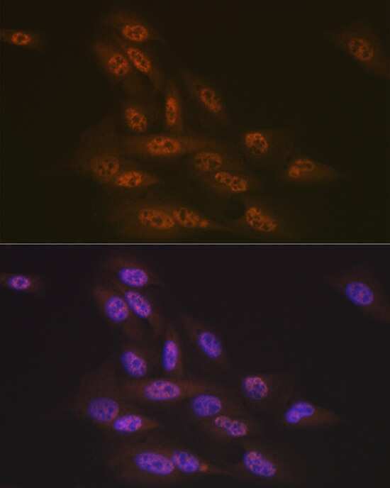
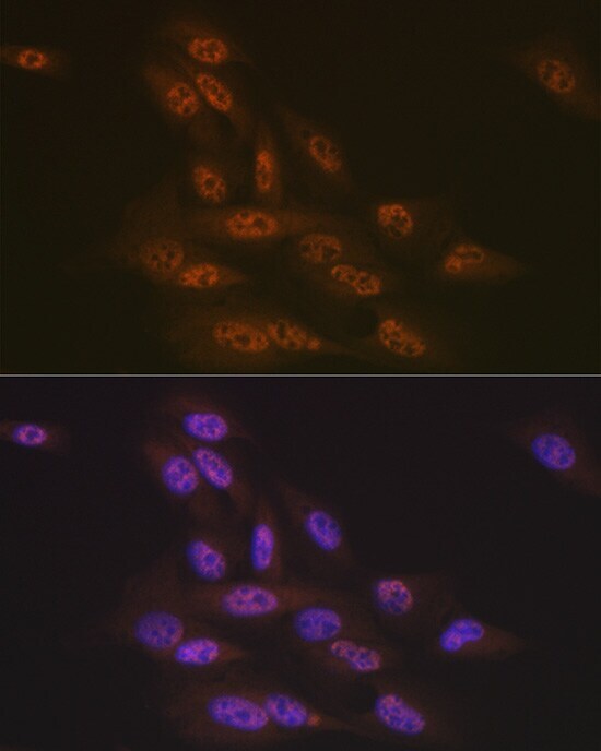
- Immunocytochemistry/Immunofluorescence analysis of TNPO3 in U-2 OS cells using TNPO3 Recombinant Monoclonal Antibody (Product # MA5-37991) at a dilution of 1:100. Blue: DAPI for nuclear staining.
- Submitted by
- Invitrogen Antibodies (provider)
- Main image

- Experimental details
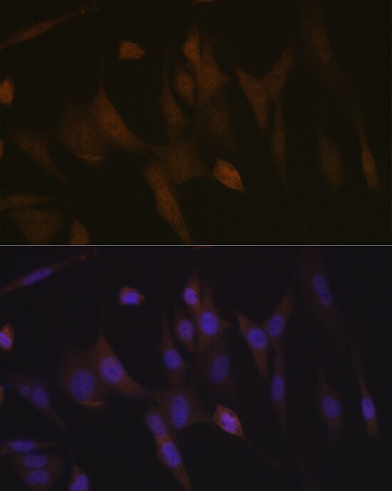
- Immunocytochemistry/Immunofluorescence analysis of TNPO3 in NIH-3T3 cells using TNPO3 Recombinant Monoclonal Antibody (Product # MA5-37991) at a dilution of 1:100. Blue: DAPI for nuclear staining.
- Submitted by
- Invitrogen Antibodies (provider)
- Main image
- Experimental details
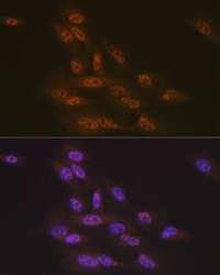
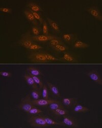
- Immunocytochemistry/Immunofluorescence analysis of TNPO3 in U-2 OS cells using TNPO3 Recombinant Monoclonal Antibody (Product # MA5-37991) at a dilution of 1:100. Blue: DAPI for nuclear staining.
- Submitted by
- Invitrogen Antibodies (provider)
- Main image
- Experimental details
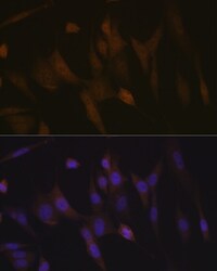
- Immunofluorescence analysis of TNPO3 in NIH-3T3 cells. Samples were incubated with TNPO3 Monoclonal antibody (Product # MA5-37991) using a dilution of 1:100 (40x lens). Blue: DAPI for nuclear staining.

- Submitted by
- Invitrogen Antibodies (provider)
- Main image

- Experimental details
- Immunofluorescence analysis of TNPO3 in U-2 OS cells. Samples were incubated with TNPO3 Monoclonal antibody (Product # MA5-37991) using a dilution of 1:100 (40x lens). Blue: DAPI for nuclear staining.

Supportive validation
- Submitted by
- Invitrogen Antibodies (provider)
- Main image
- Experimental details
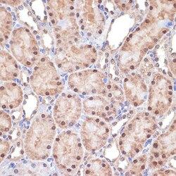
- Immunohistochemistry analysis of TNPO3 in paraffin-embedded rat kidney. Samples were incubated with TNPO3 Monoclonal antibody (Product # MA5-37991) using a dilution of 1:100 (40x lens). Perform microwave antigen retrieval with 10 mM Tris/EDTA buffer pH 9.0 before commencing with IHC staining protocol.

- Submitted by
- Invitrogen Antibodies (provider)
- Main image
- Experimental details
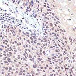
- Immunohistochemistry analysis of TNPO3 in paraffin-embedded human esophageal cancer. Samples were incubated with TNPO3 Monoclonal antibody (Product # MA5-37991) using a dilution of 1:100 (40x lens). Perform microwave antigen retrieval with 10 mM Tris/EDTA buffer pH 9.0 before commencing with IHC staining protocol.
