 Explore
 Validate
 Learn
Learn Western blot
 ELISA
ELISAAntibody data
- Antibody Data
- Antigen structure
- References [1]
- Comments [0]
- Validations
- ELISA [2]
- Immunoprecipitation [1]
- Other assay [2]
Submit
Validation data
Reference
Comment
Report error
- Product number
- MA5-19117 - Provider product page

- Provider
- Invitrogen Antibodies
- Product name
- HOXB9 Monoclonal Antibody (2E8)
- Antibody type
- Monoclonal
- Antigen
- Recombinant full-length protein
- Description
- Peptide Sequence: PLSPHASGSL PSVYHPYIQP QGVPPAESRY LRTWLEPAPR GEAAPGQGQA AVKAEPLLGA PGELLKQGTP EYSLETSAGR EAVLSNQRPG YGDNKICEG
- Reactivity
- Human
- Host
- Mouse
- Isotype
- IgG
- Antibody clone number
- 2E8
- Vial size
- 100 μg
- Concentration
- 0.5 mg/mL
- Storage
- -20°C, Avoid Freeze/Thaw Cycles
Submitted references Insights into homeobox B9: a propeller for metastasis in dormant prostate cancer progenitor cells.
Sui Y, Hu W, Zhang W, Li D, Zhu H, You Q, Zhu R, Yi Q, Tang T, Gao L, Zhu S, Yang T
British journal of cancer 2021 Sep;125(7):1003-1015
British journal of cancer 2021 Sep;125(7):1003-1015
No comments: Submit comment
Supportive validation
- Submitted by
- Invitrogen Antibodies (provider)
- Main image

- Experimental details
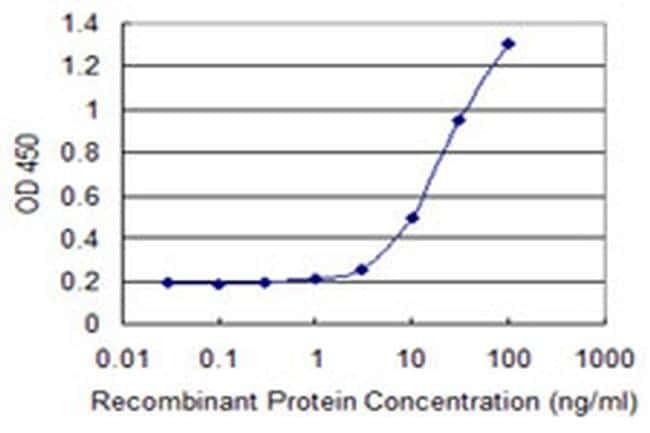
- Detection limit for recombinant GST tagged HOXB9 using anti-HOXB9 monoclonal antibody (Product # MA5-19117) is 1 ng/mL as a capture antibody.

- Submitted by
- Invitrogen Antibodies (provider)
- Main image
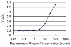
- Experimental details
- Detection limit for recombinant GST tagged HOXB9 using anti-HOXB9 monoclonal antibody (Product # MA5-19117) is 1 ng/mL as a capture antibody.
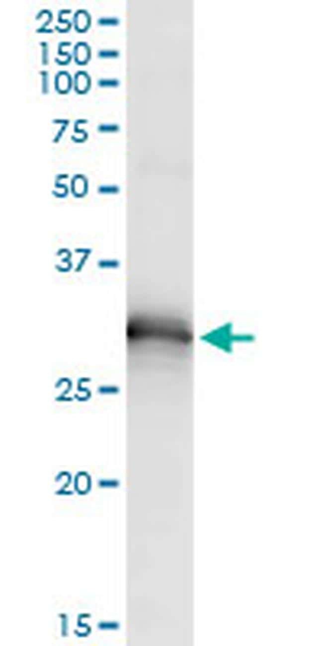
Supportive validation
- Submitted by
- Invitrogen Antibodies (provider)
- Main image

- Experimental details
- Immunoprecipitation of HOXB9 transfected lysate using anti-HOXB9 monoclonal antibody (Product # MA5-19117) and Protein A Magnetic Beads, and immunoblotted with HOXB9 rabbit polyclonal antibody.
Supportive validation
- Submitted by
- Invitrogen Antibodies (provider)
- Main image
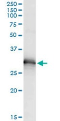
- Experimental details
- Immunoprecipitation of HOXB9 transfected lysate using anti-HOXB9 monoclonal antibody (Product # MA5-19117) and Protein A Magnetic Beads, and immunoblotted with HOXB9 rabbit polyclonal antibody.

- Submitted by
- Invitrogen Antibodies (provider)
- Main image
- Experimental details
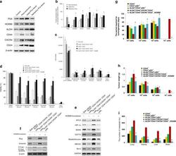
- Fig. 6 The ALDH + CD44 + CXCR4 + CD24 + subpopulation in human PCa tissues. a Western blot analysis was performed to determine the protein expressions of PSA, HOXB9, ALDH, CD44, CXCR4 and CD24 in the controls (PCa tissue with Gleason score 6), para-carcinoma (2 mm away from PCa tissue), initial PCa tissue (derived from PCa at first diagnosis via radical prostatectomy) and refractory PCa tissue (derived after recurrence), respectively. beta-actin was used as an internal control. b Quantification of ( a ). * P < 0.05 vs. initial PCa tissues. ( n = 6). c Human PCa tissue was subcutaneously implanted into NOD-SCID mice to establish a patient-derived xenograft (PDX) model. Subsets of cells (as indicated) were derived from the PDX model and seeded in 96-well plates (1 x 10 4 cells/well) and treated with different anti-androgens (as indicated) and chemotherapeutic agents, with 0.2% DMSO and 0.5% H 2 O 2 were used as negative and positive controls, respectively. After 48 h of treatment, cells were incubated with alamarBlue solution for 4 h, and cell viability was measured with excitation wavelength at 530-560 nm and emission wavelength at 590 nm using a TECAN Infinite 200 PRO microplate reader. d Subsets of cells (as indicated) were derived from the PDX model and seeded in 96-well plates (1 x 10 4 cells/well). Cells were treated with different chemotherapeutic drugs, as indicated, for 72 h. Then, a WST-1 proliferation assay was performed. The absorbance was measured at 450 nm using a
