Explore
 Validate
 Learn
Learn Western blot
Antibody data
- Antibody Data
- Antigen structure
- References [0]
- Comments [0]
- Validations
- Western blot [3]
- Immunohistochemistry [1]
- Flow cytometry [1]
Submit
Validation data
Reference
Comment
Report error
- Product number
- AGR-053-25UL - Provider product page

- Provider
- Invitrogen Antibodies
- Product name
- GPR83 (extracellular) Polyclonal Antibody
- Antibody type
- Polyclonal
- Antigen
- Other
- Reactivity
- Human, Mouse, Rat
- Host
- Rabbit
- Isotype
- IgG
- Vial size
- 25 µL
- Concentration
- 0.8 mg/mL
- Storage
- -20° C, Avoid Freeze/Thaw Cycles
No comments: Submit comment
Supportive validation
- Submitted by
- Invitrogen Antibodies (provider)
- Main image
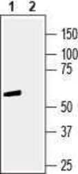
- Experimental details
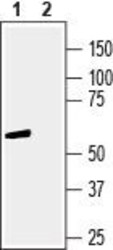
- Western blot analysis of human Jurkat T-cell leukemia cell line lysate: - 1. Anti-GPR83 (extracellular) Antibody (#AGR-053), (1:400). 2. Anti-GPR83 (extracellular) Antibody , preincubated with GPR83 (extracellular) Blocking Peptide (#BLP-GR053).
- Submitted by
- Invitrogen Antibodies (provider)
- Main image
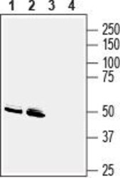
- Experimental details
- Western blot analysis of mouse brain membranes (lanes 1 and 3) and rat brain membranes (lanes 2 and 4): - 1, 2. Anti-GPR83 (extracellular) Antibody (#AGR-053), (1:400). 3, 4. Anti-GPR83 (extracellular) Antibody , preincubated with GPR83 (extracellular) Blocking Peptide (#BLP-GR053).

- Submitted by
- Invitrogen Antibodies (provider)
- Main image
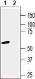
- Experimental details
- Western blot analysis of human Jurkat T-cell leukemia cell line lysate: - 1. Anti-GPR83 (extracellular) Antibody (#AGR-053), (1:400). 2. Anti-GPR83 (extracellular) Antibody , preincubated with GPR83 (extracellular) Blocking Peptide (#BLP-GR053).

Supportive validation
- Submitted by
- Invitrogen Antibodies (provider)
- Main image

- Experimental details
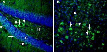
- Expression of GPR83 in mouse hippocampus and cortex - Immunohistochemical staining of perfusion-fixed frozen mouse brain sections with Anti-GPR83 (extracellular) Antibody (#AGR-053), (1:3000), followed by goat Anti-rabbit-AlexaFluor-488. A. GPR83 staining (green) in hippocampal dentate gyrus is detected in hilar interneurons (vertical arrows) and around cells of the granule layer (horizontal arrows). H = hilus of dentate gyrus. G = granule layer. B. GPR83 immunoreactivity (green) in parietal cortex is observed in neurons of the pyramidal layer (vertical arrows) and in their apical dendrites (horizontal arrows). Cell nuclei are stained with DAPI (blue).
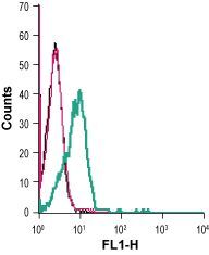
Supportive validation
- Submitted by
- Invitrogen Antibodies (provider)
- Main image
- Experimental details
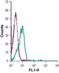
- Cell surface detection of GPR83 in live intact human Jurkat T-cell leukemia cells: - (black line) cells. (red) Cells + goat- Anti-rabbit-FITC. (green) Cells + Anti-GPR83 (extracellular) Antibody (#AGR-053), (5 µg) + goat- Anti-rabbit-FITC.