Explore
 Validate
 Learn
Learn Western blot
 Immunocytochemistry
Antibody data
- Antibody Data
- Antigen structure
- References [1]
- Comments [0]
- Validations
- Western blot [1]
Submit
Validation data
Reference
Comment
Report error
- Product number
- A00835-1 - Provider product page

- Provider
- Boster Biological Technology
- Product name
- Anti-Bile Acid Receptor NR1H4 Antibody Picoband™
- Antibody type
- Polyclonal
- Description
- Polyclonal antibody for FXR/NR1H4 detection. Host: Rabbit.Size: 100ug/vial. Tested applications: WB, ICC/IF, FCM. Reactive species: Human;Mouse. FXR/NR1H4 information: Molecular Weight: 55914 MW; Subcellular Localization: Nucleus ; Tissue Specificity: Liver and hepatocyte-related cells express mainly FXRalpha1-type isoforms with isoform 3 and isoform 4 in approximative equal proportions. In intestine and kidney mainly FXRalpha2-type isoforms are expressed with isoform 1 and isoform 2 in approximative equal proportions. Expressed in pancreatic beta cells and macrophages.
- Reactivity
- Human, Mouse
- Host
- Rabbit
- Vial size
- 100ug/vial
- Concentration
- Add 0.2ml of distilled water will yield a concentration of 500ug/ml.
- Storage
- At -20°C for one year. After reconstitution, at 4°C for one month. It can also be aliquoted and stored frozen at -20°C for a longer time. Avoid repeated freezing and thawing.
- Handling
- Add 0.2ml of distilled water will yield a concentration of 500ug/ml.
Submitted references The changes of hepatic bile acid synthesis and transport and bile acids profiles in isopsoralen-induced liver injury C57BL/6J mice.
Men WJ, Meng ZJ, Wang Q, Chen MY, Zhai YX, Shi H, Wang AH, Zhou K
Pharmaceutical biology 2022 Dec;60(1):1701-1709
Pharmaceutical biology 2022 Dec;60(1):1701-1709
No comments: Submit comment
Supportive validation
- Submitted by
- Boster Biological Technology (provider)
- Main image
- Experimental details
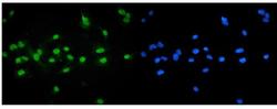
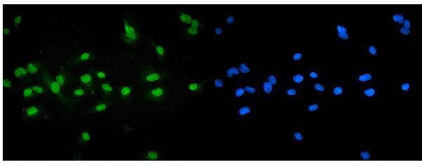
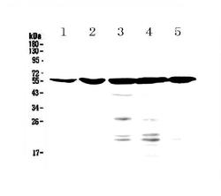
- Western blot analysis of NR1H4 using anti-NR1H4 antibody (A00835-1). Electrophoresis was performed on a 5-20% SDS-PAGE gel at 70V (Stacking gel) / 90V (Resolving gel) for 2-3 hours. The sample well of each lane was loaded with 50ug of sample under reducing conditions. Lane 1: mouse liver tissue lysate,Lane 2: mouse small intestine tissue lysate,Lane 3: human HepG2 whole cell lysate,Lane 4: human A549 whole cell lysate,Lane 5: human U2OS whole cell lysate. After Electrophoresis, proteins were transferred to a Nitrocellulose membrane at 150mA for 50-90 minutes. Blocked the membrane with 5% Non-fat Milk/ TBS for 1.5 hour at RT. The membrane was incubated with rabbit anti-NR1H4 antigen affinity purified polyclonal antibody (Catalog # A00835-1) at 0.5 μg/mL overnight at 4°C, then washed with TBS-0.1%Tween 3 times with 5 minutes each and probed with a goat anti-rabbit IgG-HRP secondary antibody at a dilution of 1:10000 for 1.5 hour at RT. The signal is developed using an Enhanced Chemiluminescent detection (ECL) kit (Catalog # EK1002) with Tanon 5200 system. A specific band was detected for NR1H4 at approximately 56KD. The expected band size for NR1H4 is at 56KD.
- Additional image