Explore
 Validate
 Learn
Learn Western blot
 Immunocytochemistry
Antibody data
- Antibody Data
- Antigen structure
- References [1]
- Comments [0]
- Validations
- Western blot [5]
- Immunocytochemistry [2]
- Immunoprecipitation [1]
Submit
Validation data
Reference
Comment
Report error
- Product number
- GTX630396 - Provider product page

- Provider
- GeneTex
- Product name
- NDP52 antibody [GT422]
- Antibody type
- Monoclonal
- Reactivity
- Human
- Host
- Mouse
Submitted references Attenuation of cGAS-STING signaling is mediated by a p62/SQSTM1-dependent autophagy pathway activated by TBK1.
Prabakaran T, Bodda C, Krapp C, Zhang BC, Christensen MH, Sun C, Reinert L, Cai Y, Jensen SB, Skouboe MK, Nyengaard JR, Thompson CB, Lebbink RJ, Sen GC, van Loo G, Nielsen R, Komatsu M, Nejsum LN, Jakobsen MR, Gyrd-Hansen M, Paludan SR
The EMBO journal 2018 Apr 13;37(8)
The EMBO journal 2018 Apr 13;37(8)
No comments: Submit comment
Enhanced validation
Supportive validation
- Submitted by
- GeneTex (provider)
- Enhanced method
- Genetic validation
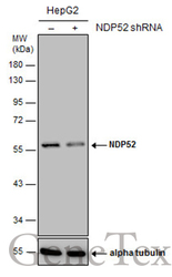
- Main image
- Experimental details
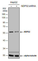
- Non-transfected (¡V) and transfected (+) HepG2 whole cell extracts (30 ?g) were separated by 10% SDS-PAGE, and the membrane was blotted with NDP52 antibody [GT422] (GTX630396) diluted at 1:1000.
Supportive validation
- Submitted by
- GeneTex (provider)
- Main image
- Experimental details
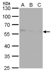
- NDP52 antibody [GT422] detects NDP52 protein by western blot analysis.A. 30 £gg Huh7 whole cell lysate/extractB. 30 £gg Hep3B whole cell lysate/extractC. 30 £gg HepG2 whole cell lysate/extract10 % SDS-PAGENDP52 antibody [GT422] (GTX630396) dilution: 1:1000

- Submitted by
- GeneTex (provider)
- Main image
- Experimental details
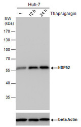
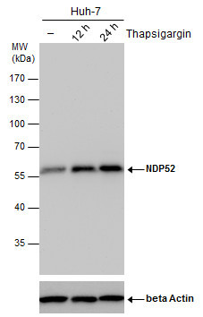
- NDP52 antibody detects NDP52 protein by western blot analysis. Un-treated (-) and treated (+, Thapsigargin treatment for 12hrs and 24hrs) Huh-7 whole cell extracts (30 £gg) were separated by 10% SDS-PAGE, and the membrane was blotted with NDP52 antibody (GTX630396) diluted by 1:500.The ACTB was used as internal control (GTX110564, 1:50000) shown at the bottom panel.
- Submitted by
- GeneTex (provider)
- Main image
- Experimental details
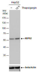
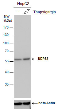
- NDP52 antibody detects NDP52 protein by western blot analysis. Un-treated (-) and treated (+, Thapsigargin treatment for 12hrs) HepG2 whole cell extracts (30 £gg) were separated by 10% SDS-PAGE, and the membrane was blotted with NDP52 antibody (GTX630396) diluted by 1:500.The ACTB was used as internal control (GTX110564, 1:50000) shown at the bottom panel.
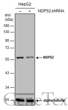
- Submitted by
- GeneTex (provider)
- Main image
- Experimental details
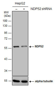
- Non-transfected (¡V) and transfected (+) HepG2 whole cell extracts (30 ?g) were separated by 10% SDS-PAGE, and the membrane was blotted with NDP52 antibody [GT422] (GTX630396) diluted at 1:1000.
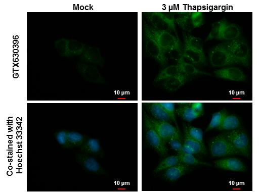
Supportive validation
- Submitted by
- GeneTex (provider)
- Main image

- Experimental details
- NDP52 antibody [GT422] detects NDP52 protein at autophagosome by immunofluorescent analysis. Samples: Hep G2 cells mock (left) and treated with 3 £gM Thapsigargin for 16 hrs (right) were fixed in ice-cold MeOH for 10 min and permeabilized with 100% MeOH for 30 sec.Green: NDP52 protein stained by NDP52 antibody [GT422] (GTX630396) diluted at 1:1000.Blue: Hoechst 33342 staining.Scale bar = 10 £gm.
- Submitted by
- GeneTex (provider)
- Main image

- Experimental details
- NDP52 antibody [GT422] detects NDP52 protein at autophagosome by immunofluorescent analysis. Samples: HeLa cells mock (left) and treated with 50£gM Chloroquine for 24 hr (right) were fixed in 4% paraformaldehyde at RT for 15 min.Green: NDP52 protein stained by NDP52 antibody [GT422] (GTX630396) diluted at 1:1000.Red: Phalloidin, a F-actin marker.
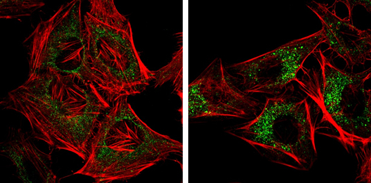
Supportive validation
- Submitted by
- GeneTex (provider)
- Main image
- Experimental details
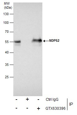
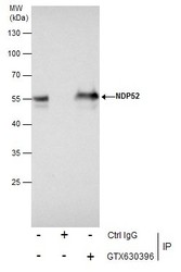
- Immunoprecipitation of NDP52 protein from Jurkat whole cell extracts using 5 £gg of NDP52 antibody [GT422] (GTX630396).Western blot analysis was performed using NDP52 antibody [GT422] (GTX630396).EasyBlot anti-Mouse IgG (GTX221667-01) was used as a secondary reagent.