Explore
 Validate
 Learn
Learn Western blot
 Immunocytochemistry
Antibody data
- Antibody Data
- Antigen structure
- References [3]
- Comments [0]
- Validations
- Western blot [7]
- Immunocytochemistry [2]
- Immunoprecipitation [1]
- Immunohistochemistry [1]
Submit
Validation data
Reference
Comment
Report error
- Product number
- GTX115378 - Provider product page

- Provider
- GeneTex
- Proper citation
- GeneTex Cat#GTX115378, RRID:AB_10620266
- Product name
- NDP52 antibody
- Antibody type
- Polyclonal
- Reactivity
- Human, Mouse
- Host
- Rabbit
Submitted references Dual Inhibition of PIK3C3 and FGFR as a New Therapeutic Approach to Treat Bladder Cancer.
Age-dependent changes in synaptic plasticity enhance tau oligomerization in the mouse hippocampus.
NDP52 activates nuclear myosin VI to enhance RNA polymerase II transcription.
Chen CH, Changou CA, Hsieh TH, Lee YC, Chu CY, Hsu KC, Wang HC, Lin YC, Lo YN, Liu YR, Liou JP, Yen Y
Clinical cancer research : an official journal of the American Association for Cancer Research 2018 Mar 1;24(5):1176-1189
Clinical cancer research : an official journal of the American Association for Cancer Research 2018 Mar 1;24(5):1176-1189
Age-dependent changes in synaptic plasticity enhance tau oligomerization in the mouse hippocampus.
Kimura T, Suzuki M, Akagi T
Acta neuropathologica communications 2017 Sep 6;5(1):67
Acta neuropathologica communications 2017 Sep 6;5(1):67
NDP52 activates nuclear myosin VI to enhance RNA polymerase II transcription.
Fili N, Hari-Gupta Y, Dos Santos Á, Cook A, Poland S, Ameer-Beg SM, Parsons M, Toseland CP
Nature communications 2017 Nov 30;8(1):1871
Nature communications 2017 Nov 30;8(1):1871
No comments: Submit comment
Enhanced validation
Supportive validation
- Submitted by
- GeneTex (provider)
- Enhanced method
- Genetic validation
- Main image
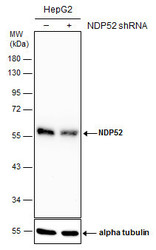
- Experimental details
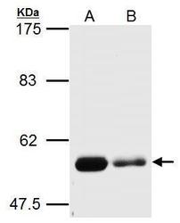
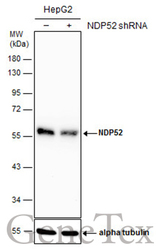
- Non-transfected (¡V) and transfected (+) HepG2 whole cell extracts (30 ?g) were separated by 10% SDS-PAGE, and the membrane was blotted with NDP52 antibody (GTX115378) diluted at 1:4000. The HRP-conjugated anti-rabbit IgG antibody (GTX213110-01) was used to detect the primary antibody.
Supportive validation
- Submitted by
- GeneTex (provider)
- Main image
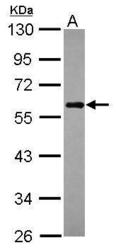
- Experimental details
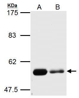
- Sample (30 ?g of whole cell lysate) A: Raji 10% SDS PAGE GTX115378 diluted at 1:1000 The HRP-conjugated anti-rabbit IgG antibody (GTX213110-01) was used to detect the primary antibody.

- Submitted by
- GeneTex (provider)
- Main image
- Experimental details
- NDP52 antibody detects NDP52 protein by Western blot analysis.A. 20 ?g Huh7 whole cell lysate/extract (untreated) B. 20 ?g Huh7 whole cell lysate/extract (3uM-Thapsipargin treatment for 12hr)0 % SDS-PAGENDP52 antibody (GTX115378) dilution: 1:1500
- Validation comment
- WB
- Submitted by
- GeneTex (provider)
- Main image
- Experimental details
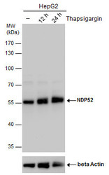
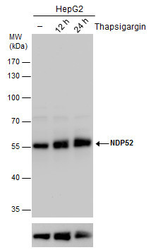
- NDP52 antibody detects NDP52 protein by western blot analysis. Un-treated (-) and treated (+, Thapsigargin treatment for 12hrs and 24hrs) HepG2 whole cell extracts (30 ?g) were separated by 10% SDS-PAGE, and the membrane was blotted with NDP52 antibody (GTX115378) diluted by 1:2000.The ACTB was used as internal control (GTX110564, 1:50000) shown at the bottom panel. The HRP-conjugated anti-rabbit IgG antibody (GTX213110-01) was used to detect the primary antibody.
- Submitted by
- GeneTex (provider)
- Main image
- Experimental details
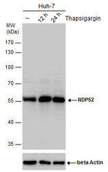
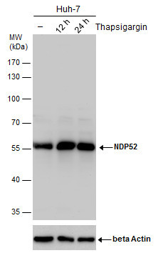
- NDP52 antibody detects NDP52 protein by western blot analysis. Un-treated (-) and treated (+, Thapsigargin treatment for 12hrs and 24hrs) Huh-7 whole cell extracts (30 ?g) were separated by 10% SDS-PAGE, and the membrane was blotted with NDP52 antibody (GTX115378) diluted by 1:2000.The ACTB was used as internal control (GTX110564, 1:50000) shown at the bottom panel. The HRP-conjugated anti-rabbit IgG antibody (GTX213110-01) was used to detect the primary antibody.
- Submitted by
- GeneTex (provider)
- Main image
- Experimental details
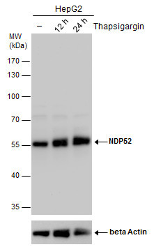
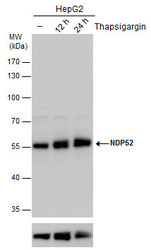
- NDP52 antibody detects NDP52 protein by western blot analysis. Un-treated (-) and treated (+, Thapsigargin treatment for 12hrs and 24hrs) HepG2 whole cell extracts (30 ?g) were separated by 10% SDS-PAGE, and the membrane was blotted with NDP52 antibody (GTX115378) diluted by 1:2000.The ACTB was used as internal control (GTX110564, 1:50000) shown at the bottom panel.
- Validation comment
- WB
- Submitted by
- GeneTex (provider)
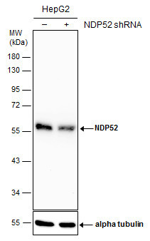
- Main image
- Experimental details
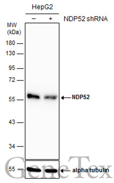
- Non-transfected (¡V) and transfected (+) HepG2 whole cell extracts (30 ?g) were separated by 10% SDS-PAGE, and the membrane was blotted with NDP52 antibody (GTX115378) diluted at 1:4000. The HRP-conjugated anti-rabbit IgG antibody (GTX213110-01) was used to detect the primary antibody.
Supportive validation
- Submitted by
- GeneTex (provider)
- Main image
- Experimental details
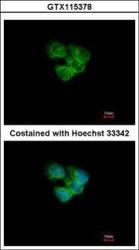
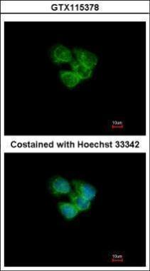
- Immunofluorescence analysis of methanol-fixed A431, using NDP52(GTX115378) antibody at 1:200 dilution.
- Submitted by
- GeneTex (provider)
- Main image

- Experimental details
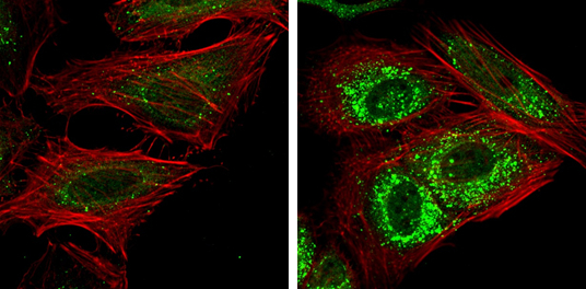
- NDP52 antibody detects NDP52 protein at autophagosome by immunofluorescent analysis. Samples: HeLa cells mock (left) and treated with 50£gM Chloroquine for 24 hr (right) were fixed in 4% paraformaldehyde at RT for 15 min.Green: NDP52 protein stained by NDP52 antibody (GTX115378) diluted at 1:1000.Red: Phalloidin, a F-actin marker.
Supportive validation
- Submitted by
- GeneTex (provider)
- Main image
- Experimental details
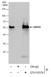
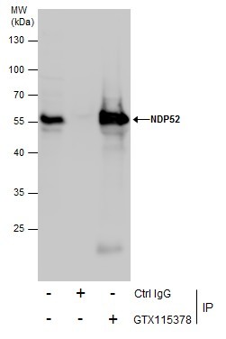
- Immunoprecipitation of NDP52 protein from Jurkat whole cell extracts using 5 £gg of NDP52 antibody (GTX115378).Western blot analysis was performed using NDP52 antibody (GTX115378).EasyBlot anti-Rabbit IgG (GTX221666-01) was used as a secondary reagent.
Supportive validation
- Submitted by
- GeneTex (provider)
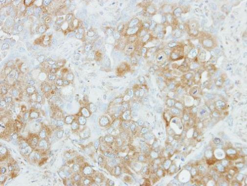
- Main image

- Experimental details
- Immunohistochemical analysis of paraffin-embedded H441 xenograft, using NDP52(GTX115378) antibody at 1:100 dilution.