Explore
 Validate
 Learn
Learn Western blot
 Immunocytochemistry
Antibody data
- Antibody Data
- Antigen structure
- References [0]
- Comments [0]
- Validations
- Immunocytochemistry [3]
- Immunohistochemistry [1]
- Other assay [1]
Submit
Validation data
Reference
Comment
Report error
- Product number
- PA5-30367 - Provider product page

- Provider
- Invitrogen Antibodies
- Product name
- CALCOCO2 Polyclonal Antibody
- Antibody type
- Polyclonal
- Antigen
- Recombinant full-length protein
- Description
- Recommended positive controls: Raji, HepG2, HepG2 whole cell extract (untreated), HepG2 whole cell extract (3 µM Thapsigargin treatment for 12 hr), HepG2 whole cell extract (3 µM Thapsigargin treatment for 24 hr), Huh-7 (untreated), Huh-7 (3 µM Thapsigargin treatment for 12 hr), Huh-7 (3 µM Thapsigargin treatment for 24 hr). Predicted reactivity: Human (99%), Chimpanzee (97%). Store product as a concentrated solution. Centrifuge briefly prior to opening the vial.
- Reactivity
- Human, Mouse
- Host
- Rabbit
- Isotype
- IgG
- Vial size
- 100 μL
- Concentration
- 1.57 mg/mL
- Storage
- Store at 4°C short term. For long term storage, store at -20°C, avoiding freeze/thaw cycles.
No comments: Submit comment
Supportive validation
- Submitted by
- Invitrogen Antibodies (provider)
- Main image
- Experimental details
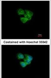
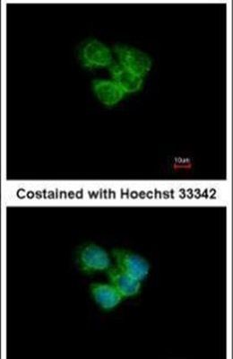
- Immunofluorescent analysis of CALCOCO2 in methanol-fixed A431 cells using a CALCOCO2 polyclonal antibody (Product # PA5-30367) at a 1:200 dilution.
- Submitted by
- Invitrogen Antibodies (provider)
- Main image

- Experimental details
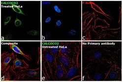
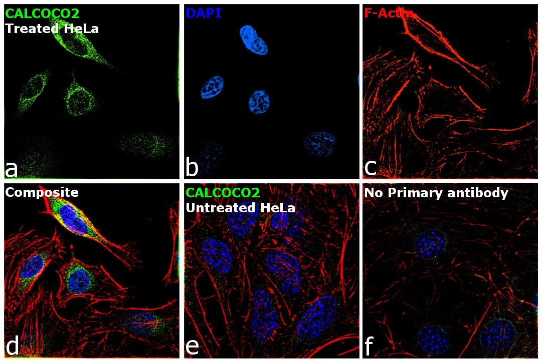
- Immunofluorescence analysis of CALCOCO2 was performed using 70% confluent log phase HeLa cells treated with 3 Thapsigargin 3uM for 48 hrs. The cells were fixed with 4% paraformaldehyde for 10 minutes, permeabilized with 0.1% Triton™ X-100 for 15 minutes, and blocked with 2% BSA for 45 minutes at room temperature. The cells were labeled with CALCOCO2 Polyclonal Antibody (Product # PA5-30367) at 1:100 in 0.1% BSA, incubated at 4 degree celsius overnight and then labeled with Goat anti-Rabbit IgG (H+L) Highly Cross-Adsorbed Secondary Antibody, Alexa Fluor Plus 488 (Product # A32731), (1:2000), for 45 minutes at room temperature (Panel a: Green). Nuclei (Panel b: Blue) were stained with ProLong™ Diamond Antifade Mountant with DAPI (Product # P36962). F-actin (Panel c: Red) was stained with Rhodamine Phalloidin (Product # R415, 1:300). Panel d represents the merged image showing Cytosol localization. Panel e represents untreated HeLa cells showing no signal Panel f represents control cells with no primary antibody to assess background. The images were captured at 60X magnification.

- Submitted by
- Invitrogen Antibodies (provider)
- Main image
- Experimental details
- Immunofluorescence analysis of CALCOCO2 was performed using 70% confluent log phase HeLa cells treated with 3 Thapsigargin 3uM for 48 hrs. The cells were fixed with 4% paraformaldehyde for 10 minutes, permeabilized with 0.1% Triton™ X-100 for 15 minutes, and blocked with 2% BSA for 45 minutes at room temperature. The cells were labeled with CALCOCO2 Polyclonal Antibody (Product # PA5-30367) at 1:100 in 0.1% BSA, incubated at 4 degree celsius overnight and then labeled with Goat anti-Rabbit IgG (H+L) Highly Cross-Adsorbed Secondary Antibody, Alexa Fluor Plus 488 (Product # A32731), (1:2000), for 45 minutes at room temperature (Panel a: Green). Nuclei (Panel b: Blue) were stained with ProLong™ Diamond Antifade Mountant with DAPI (Product # P36962). F-actin (Panel c: Red) was stained with Rhodamine Phalloidin (Product # R415, 1:300). Panel d represents the merged image showing Cytosol localization. Panel e represents untreated HeLa cells showing no signal Panel f represents control cells with no primary antibody to assess background. The images were captured at 60X magnification.
Supportive validation
- Submitted by
- Invitrogen Antibodies (provider)
- Main image

- Experimental details
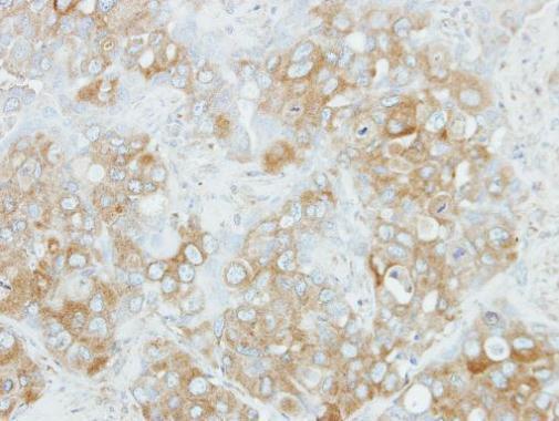
- Immunohistochemical analysis of paraffin-embedded H441 xenograft, using NDP52 (Product # PA5-30367) antibody at 1:100 dilution. Antigen Retrieval: EDTA based buffer, pH 8.0, 15 min.
Supportive validation
- Submitted by
- Invitrogen Antibodies (provider)
- Main image

- Experimental details
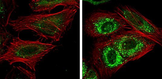
- Immunofluorescent analysis of CALCOCO2 showing staining in the autophagosome of HeLa cells. HeLa cells mock (left) and treated with 50µM Chloroquine for 24 hr (right) were fixed in 4% paraformaldehyde at RT for 15 min and stained using a CALCOCO2 polyclonal antibody (Product # PA5-30367) diluted at 1:1000. Red: Phalloidin, a F-actin marker.