Explore
 Validate
 Learn
Learn Immunocytochemistry
Antibody data
- Antibody Data
- Antigen structure
- References [1]
- Comments [0]
- Validations
- Immunocytochemistry [1]
- Chromatin Immunoprecipitation [2]
- Other assay [1]
Submit
Validation data
Reference
Comment
Report error
- Product number
- 702055 - Provider product page

- Provider
- Invitrogen Antibodies
- Product name
- TBX3 Recombinant Rabbit Monoclonal Antibody (22H23L21)
- Antibody type
- Monoclonal
- Antigen
- Other
- Description
- This antibody is predicted to react with Monkey, Pig, Rat and Mouse Recombinant rabbit monoclonal antibodies are produced using in vitro expression systems. The expression systems are developed by cloning in the specific antibody DNA sequences from immunoreactive rabbits. Then, individual clones are screened to select the best candidates for production. The advantages of using recombinant rabbit monoclonal antibodies include: better specificity and sensitivity, lot-to-lot consistency, animal origin-free formulations, and broader immunoreactivity to diverse targets due to larger rabbit immune repertoire.
- Reactivity
- Human
- Host
- Rabbit
- Isotype
- IgG
- Antibody clone number
- 22H23L21
- Vial size
- 100 μg
- Concentration
- 0.5 mg/mL
- Storage
- Store at 4°C short term. For long term storage, store at -20°C, avoiding freeze/thaw cycles.
Submitted references NODAL inhibition promotes differentiation of pacemaker-like cardiomyocytes from human induced pluripotent stem cells.
Yechikov S, Kao HKJ, Chang CW, Pretto D, Zhang XD, Sun YH, Smithers R, Sirish P, Nolta JA, Chan JW, Chiamvimonvat N, Lieu DK
Stem cell research 2020 Dec;49:102043
Stem cell research 2020 Dec;49:102043
No comments: Submit comment
Supportive validation
- Submitted by
- Invitrogen Antibodies (provider)
- Main image
- Experimental details
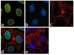
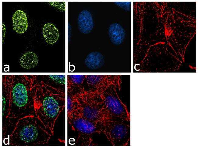
- For immunofluorescence analysis, MCF-7 cells were fixed and permeabilized for detection of endogenous TBX3 using Anti- TBX3 Recombinant Rabbit Monoclonal Antibody (Product # 702055, 2 µg/mL) and labeled with Goat anti-Rabbit IgG (Heavy Chain) Superclonal Secondary Antibody, Alexa Fluor® 488 conjugate (Product # A27034, 1:2000). Panel a) shows representative cells that were stained for detection and localization of TBX3 protein (green), Panel b) is stained for nuclei (blue) using SlowFade® Gold Antifade Mountant with DAPI (Product # S36938). Panel c) represents cytoskeletal F-actin staining using Alexa Fluor® 555 Rhodamine Phalloidin (Product # R415, 1:300). Panel d) is a composite image of Panels a, b and c clearly demonstrating nuclear localization of TBX3. Panel e) represents control cells with no primary antibody to assess background. The images were captured at 60X magnification.
Supportive validation
- Submitted by
- Invitrogen Antibodies (provider)
- Main image
- Experimental details
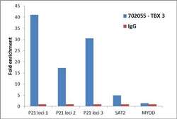
- Enrichment of endogenous TBX3 protein at specific gene loci using Anti-TBX3 Recombinant Rabbit Monoclonal Antibody: Chromatin Immunoprecipitation (ChIP) was performed using Anti-TBX3 Recombinant Rabbit Monoclonal Antibody (Product # 702055, 5 µg) on sheared chromatin from 2 million HEPG2 cells using the "MAGnify ChIP system" kit (Product # 49-2024). Normal Rabbit IgG (1 µg) was used as a negative IP control. The purified DNA was analyzed by 7500 Fast qPCR system (Product # 4351106) with optimized PCR primer pairs for the different loci of the active P21 promoter region used as positive control target genes, and the region of the inactive MYOD, SAT2 satellite repeat, used as negative control target gene. Data is presented as fold enrichment of the antibody signal versus the negative control IgG using the comparative CT method.

- Submitted by
- Invitrogen Antibodies (provider)
- Main image

- Experimental details
- Enrichment of endogenous TBX3 protein at specific gene loci using Anti-TBX3 Recombinant Rabbit Monoclonal Antibody: Chromatin Immunoprecipitation (ChIP) was performed using Anti-TBX3 Recombinant Rabbit Monoclonal Antibody (Product # 702055, 5 µg) on sheared chromatin from 2 million HEPG2 cells using the "MAGnify ChIP system" kit (Product # 49-2024). Normal Rabbit IgG (1 µg) was used as a negative IP control. The purified DNA was analyzed by 7500 Fast qPCR system (Product # 4351106) with optimized PCR primer pairs for the different loci of the active P21 promoter region used as positive control target genes, and the region of the inactive MYOD, SAT2 satellite repeat, used as negative control target gene. Data is presented as fold enrichment of the antibody signal versus the negative control IgG using the comparative CT method.

Supportive validation
- Submitted by
- Invitrogen Antibodies (provider)
- Main image
- Experimental details
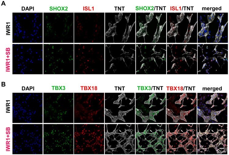
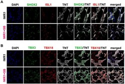
- Fig. 3. Morphology and protein expression of hiPSC-CMs differentiated by IWR1+SB. A) SHOX2 and ISL1 staining in hiPSC-CMs of 14 days post-differentiation that were differentiated by the addition of IWR1+SB from day 3-5 compared to the IWR1 control. B) TBX3 and TBX18 staining in hiPSC-CMs from the IWR1+SB-differentiation relative to the IWR1 control. DAPI counterstain and TNT staining were included in all stained samples to allow identification of all cells and cardiomyocytes, respectively.