 Explore
 Validate
 Learn
Learn Western blot
 Immunocytochemistry
Antibody data
- Antibody Data
- Antigen structure
- References [3]
- Comments [0]
- Validations
- Immunocytochemistry [1]
- Flow cytometry [1]
- Other assay [2]
Submit
Validation data
Reference
Comment
Report error
- Product number
- PA5-35373 - Provider product page

- Provider
- Invitrogen Antibodies
- Product name
- EMX1 Polyclonal Antibody
- Antibody type
- Polyclonal
- Antigen
- Synthetic peptide
- Reactivity
- Human, Mouse
- Host
- Rabbit
- Isotype
- IgG
- Vial size
- 400 μL
- Concentration
- 2 mg/mL
- Storage
- Store at 4°C short term. For long term storage, store at -20°C, avoiding freeze/thaw cycles.
Submitted references The eIF4E homolog 4EHP (eIF4E2) regulates hippocampal long-term depression and impacts social behavior.
16p11.2 microdeletion imparts transcriptional alterations in human iPSC-derived models of early neural development.
Digenic Inheritance of PROKR2 and WDR11 Mutations in Pituitary Stalk Interruption Syndrome.
Wiebe S, Meng XQ, Kim SH, Zhang X, Lacaille JC, Aguilar-Valles A, Sonenberg N
Molecular autism 2020 Nov 23;11(1):92
Molecular autism 2020 Nov 23;11(1):92
16p11.2 microdeletion imparts transcriptional alterations in human iPSC-derived models of early neural development.
Roth JG, Muench KL, Asokan A, Mallett VM, Gai H, Verma Y, Weber S, Charlton C, Fowler JL, Loh KM, Dolmetsch RE, Palmer TD
eLife 2020 Nov 10;9
eLife 2020 Nov 10;9
Digenic Inheritance of PROKR2 and WDR11 Mutations in Pituitary Stalk Interruption Syndrome.
McCormack SE, Li D, Kim YJ, Lee JY, Kim SH, Rapaport R, Levine MA
The Journal of clinical endocrinology and metabolism 2017 Jul 1;102(7):2501-2507
The Journal of clinical endocrinology and metabolism 2017 Jul 1;102(7):2501-2507
No comments: Submit comment
Supportive validation
- Submitted by
- Invitrogen Antibodies (provider)
- Main image
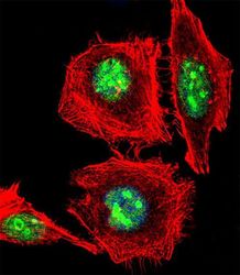
- Experimental details
- Immunofluorescent analysis of EMX1 showing staining in the nucleus of Hela cells using an EMX1 polyclonal antibody (Product # PA5-35373) followed by detection using a fluorescent conjugated secondary antibody (green). Cytoplasmic actin was stained with a fluorescent red phalloidin (7units/ml, 1 h at 37°C). Nuclei were stained with DAPI (blue) (10 µg/mL, 10 min).
Supportive validation
- Submitted by
- Invitrogen Antibodies (provider)
- Main image
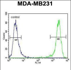
- Experimental details
- Flow cytometry analysis of EMX1 in MDA-MB231 cells (right) compared to a negative control (left) using an EMX1 polyclonal antibody (Product # PA5-35373) followed by detection using a FITC-conjugated goat-anti-rabbit secondary antibody.
Supportive validation
- Submitted by
- Invitrogen Antibodies (provider)
- Main image

- Experimental details
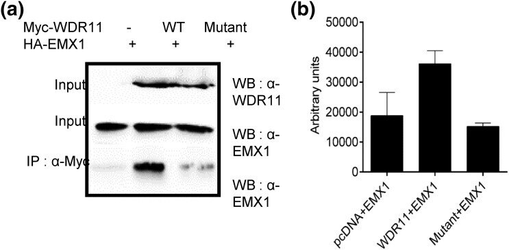
- Figure 2. Mutant WDR11 fails to bind to EMX1. (a) WDR11 fusion proteins containing a Myc epitope tag WT or the c.1306A>G variant (mutant) were coexpressed in HEK293 cells along with HA-EMX1 protein. The total cell lysates were immunoprecipitated with anti-Myc antibody, and the association of EMX1 protein was determined by immunoblot analysis using anti-EMX1 antibody. Empty pcDNA vector (-) was included as a negative control. (b) The average densitometry values of the EMX1 band intensity obtained from three independent experiments are shown with the standard deviations (error bars).
- Submitted by
- Invitrogen Antibodies (provider)
- Main image
- Experimental details
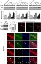
- Fig. 1 4EHP expression in the brain. a - c Developmental expression of 4EHP, GIGYF2, and GAPDH in the cortex, hippocampus, and cerebellum, respectively, as measured by western blot. Quantification of a, b, and c (lower panel, n = 3 per group, normalized to the average of all age points for each protein per membrane). d 4EHP expression in a synaptosome preparation (left panel). PSD95 was enriched in the synaptosome (Syn) as opposed to the cytosol (Cyto), demonstrating proper synaptosome preparation. GAPDH and beta-actin were used as loading controls. Quantification of d (right panel, n = 3). e Primary neurons were derived from the hippocampus of wild-type mice and cultured for 14 d. Immunofluorescent analysis confirmed 4EHP expression in the synapse by colocalization with PSD95 (merge). The scale bar represents 20 um in the upper panel of images and 5 um in the lower panel of images. The lower panel of images correspond to 4 x zoom of the upper panel of images defined by the white box. Analysis of cell-type-specific expression of 4EHP by colocalization with f Empty Spiracles Homeobox 1 (EMX1, defining excitatory neurons), g parvalbumin (PV, defining a subset of inhibitory neurons), h somatostatin (SST, defining another subset of inhibitory neurons), and I laminin (LAMA1, defining endothelial cells) in the hippocampus of wild-type mice. 4EHP expression is colored in red, the cell type marker in green, and Hoechst-stained nucleus in blue. Arrows indicate a positive signal for th
