Explore
 Validate
 Learn
Learn Western blot
Antibody data
- Antibody Data
- Antigen structure
- References [1]
- Comments [0]
- Validations
- Western blot [1]
- Immunocytochemistry [1]
- Other assay [1]
Submit
Validation data
Reference
Comment
Report error
- Product number
- PA5-101636 - Provider product page

- Provider
- Invitrogen Antibodies
- Product name
- EMX1 Polyclonal Antibody
- Antibody type
- Polyclonal
- Antigen
- Synthetic peptide
- Description
- Antibody detects endogenous levels of total EMX1.
- Reactivity
- Human, Mouse, Rat
- Host
- Rabbit
- Isotype
- IgG
- Vial size
- 100 μL
- Concentration
- 1 mg/mL
- Storage
- -20°C
Submitted references EMX1 functions as a tumor inhibitor in spinal cord glioma through transcriptional suppression of WASF2 and inactivation of the Wnt/β-catenin axis.
Han Z, Mou Z, Jing Y, Jiang R, Sun T
Brain and behavior 2022 Aug;12(8):e2684
Brain and behavior 2022 Aug;12(8):e2684
No comments: Submit comment
Supportive validation
- Submitted by
- Invitrogen Antibodies (provider)
- Main image
- Experimental details
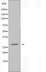
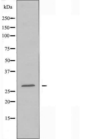
- Western blot analysis of EMX1 in HUVEC cell lysate. Samples were incubated with EMX1 polyclonal antibody (Product # PA5-101636).
Supportive validation
- Submitted by
- Invitrogen Antibodies (provider)
- Main image
- Experimental details
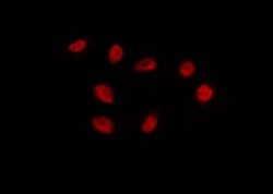
- Immunofluorescent analysis of EMX1 in HepG2. Samples were fixed with paraformaldehyde, permeabilized with 0.1% Triton X-100, blocked with 10% serum (45 min at 25°C) incubated with EMX1 polyclonal antibody (Product # PA5-101636) using a dilution of 1:200 (1 hr, 37°C), and followed by goat anti-rabbit IgG Alexa Fluor 594 at a dilution of 1:600.

Supportive validation
- Submitted by
- Invitrogen Antibodies (provider)
- Main image
- Experimental details
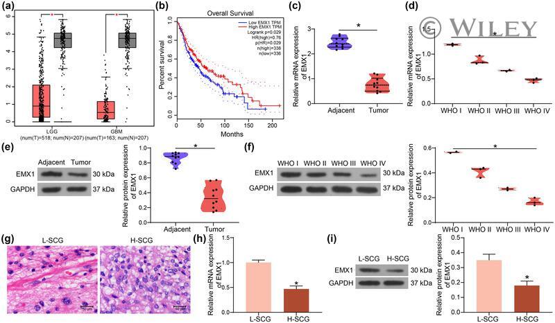
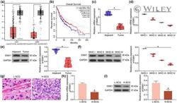
- 1 FIGURE Poor expression of EMX1 in SCG is correlated with increased tumor grades. (a) expression of EMX1 in glioma patients in the GEPIA database; (b) correlation between EMX1 expression and the overall survival of glioma patients in the GEPIA database; (c) expression of EMX1 mRNA in the SCG tissues and the adjacent normal tissues examined by RT-qPCR; (d) expression of EMX1 mRNA in different grades of SCG; (e) expression of EMX1 protein in the SCG tissues and the adjacent normal tissues examined by western blot analysis; (f) expression of EMX1 protein in different grades of SCG; (g) histological characteristics of L-SCG and H-SCG tissues observed by HE staining; H-I, mRNA (h) and protein (i) levels of EMX1 in L-SCG and H-SCG cells detected by RT-qPCR and western blot assays. Three repetitions were performed for cellular experiments. Differences were analyzed by the paired t test (c and e), the unpaired t test (h and i), and one-way ANOVA (d and f). * p < .05