 Explore
 Validate
 Learn
Learn Western blot
Antibody data
- Antibody Data
- Antigen structure
- References [0]
- Comments [0]
- Validations
- Western blot [6]
- Immunohistochemistry [3]
Submit
Validation data
Reference
Comment
Report error
- Product number
- PA5-40912 - Provider product page

- Provider
- Invitrogen Antibodies
- Product name
- NRF1 Polyclonal Antibody
- Antibody type
- Polyclonal
- Antigen
- Synthetic peptide
- Description
- Peptide sequence: WATLQGGEMT IQTTQASEAT QAVASLAEAA VAASQEMQQG ATVTMALNSE
- Concentration
- 0.5 mg/mL
No comments: Submit comment
Supportive validation
- Submitted by
- Invitrogen Antibodies (provider)
- Main image
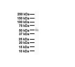
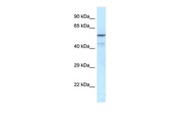
- Experimental details
- Western blot analysis of human heart cells using an anti-NRF1 polyclonal antibody (Product # PA5-40912).
- Submitted by
- Invitrogen Antibodies (provider)
- Main image

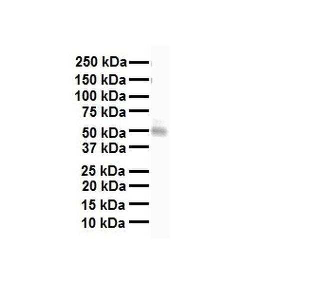
- Experimental details
- Western blot analysis of human 293T cells using an anti-NRF1 polyclonal antibody (Product # PA5-40912).

- Submitted by
- Invitrogen Antibodies (provider)
- Main image
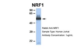
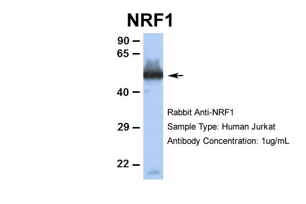
- Experimental details
- Western blot analysis of human Jurkat cells using an anti-NRF1 polyclonal antibody (Product # PA5-40912).
- Submitted by
- Invitrogen Antibodies (provider)
- Main image
- Experimental details
- Western blot analysis of transfected 293T cells using an anti-NRF1 polyclonal antibody (Product # PA5-40912).

- Submitted by
- Invitrogen Antibodies (provider)
- Main image

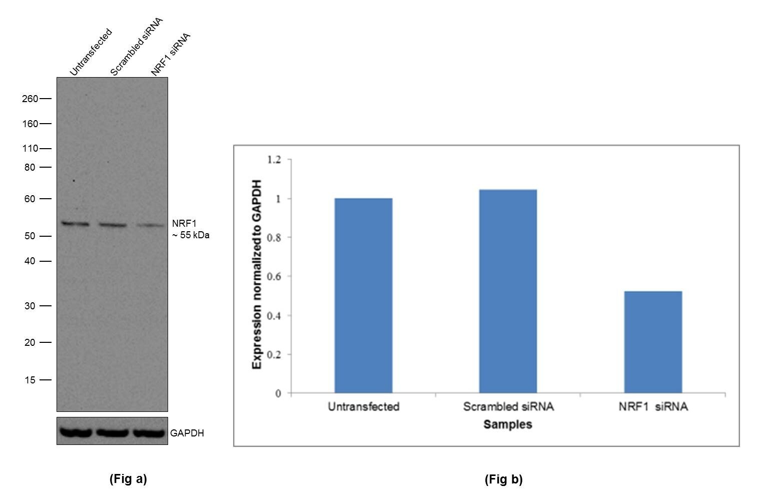
- Experimental details
- KD of NRF1 was achieved by transfecting HeLa cells with NRF1 specific siRNAs (Silencer® select Product # s32459, s32460). Western blot analysis (Fig. a) was performed using Whole cell extracts from the NRF1 KD cells (lane 3), non-specific scrambled siRNA transfected cells (lane 2) and untransfected cells (lane 1). The blot was probed with NRF1 Polyclonal Antibody (Product # PA5-40912, 0.5 µg/mL) and Goat Anti-Rabbit IgG Secondary Antibody, HRP conjugate (Product # A27036, 1:4000 dilution). Densitometric analysis of this western blot is shown in histogram (Fig. b). Decrease in signal upon siRNA mediated knock down confirms that antibody is specific to NRF1. .
- Submitted by
- Invitrogen Antibodies (provider)
- Main image

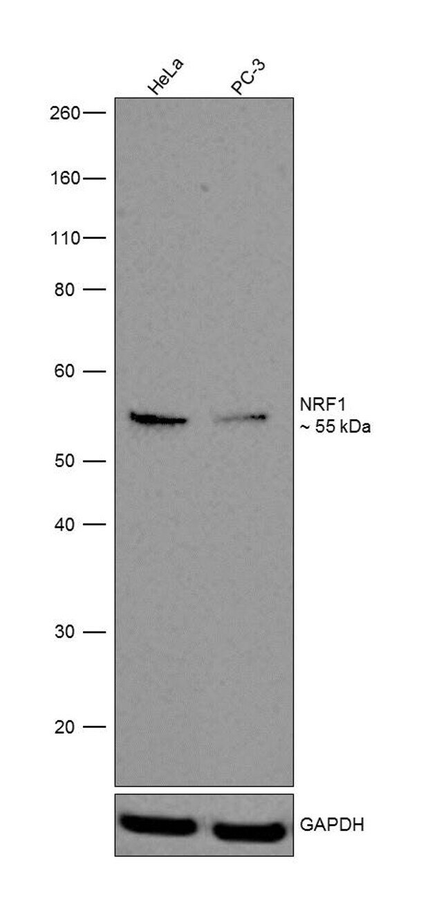
- Experimental details
- Western blot was performed using Anti-NRF1 Rabbit Polyclonal Antibody (Product # PA5-40912). A 55 kDa band corresponding to NRF1 was observed across cell lines tested. Modified whole cell extracts (1% SDS) (30 µg lysate) of HeLa (Lane 1) and PC-3 (Lane 2) were electrophoresed using Novex® NuPAGE® 4-12 % Bis-Tris gel (Product # NP0322BOX). Resolved proteins were then transferred onto a nitrocellulose membrane (Product # IB23001) by iBlot® 2 Dry Blotting System (Product # IB21001). The blot was probed with the primary antibody (0.5 µg/mL) and detected by Goat Anti-Rabbit IgG Secondary Antibody, HRP conjugate (Product # A27036, 1:4000 dilution) using the iBright FL 1000 (Product # A32752). Chemiluminescent detection was performed using Novex® ECL Chemiluminescent Substrate Reagent Kit (Product # WP20005).
Supportive validation
- Submitted by
- Invitrogen Antibodies (provider)
- Main image

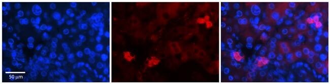
- Experimental details
- Immunohistochemistry (paraffin-embedded) analysis of human adult liver tissue using an anti-NRF1 polyclonal antibody (Product # PA5-40912).
- Submitted by
- Invitrogen Antibodies (provider)
- Main image
- Experimental details
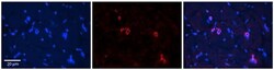
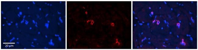
- Immunohistochemistry (paraffin-embedded) analysis of human adult heart tissue using an anti-NRF1 polyclonal antibody (Product # PA5-40912).
- Submitted by
- Invitrogen Antibodies (provider)
- Main image
- Experimental details
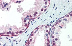
- Immunohistochemistry (paraffin-embedded) analysis of human prostate tissue using an anti-NRF1 polyclonal antibody (Product # PA5-40912).
